Review
. 2014 Jun 12:2:749-54.
doi: 10.1016/j.redox.2014.06.004. eCollection 2014.

Variants of mitochondrial autophagy: Types 1 and 2 mitophagy and micromitophagy (Type 3)

Affiliations
Review

Variants of mitochondrial autophagy: Types 1 and 2 mitophagy and micromitophagy (Type 3)

John J Lemasters. Redox Biol. .

Abstract

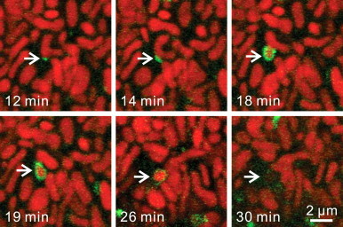
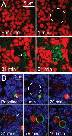
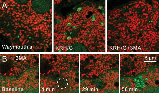
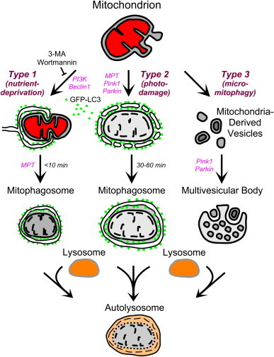
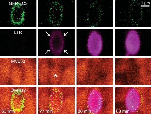
Mitophagy (mitochondrial autophagy), which removes damaged, effete and superfluous mitochondria, has several distinct variants. In Type 1 mitophagy occurring during nutrient deprivation, preautophagic structures (PAS) grow into cup-shaped phagophores that surround and sequester individual mitochondria into mitophagosomes, a process requiring phosphatidylinositol-3-kinase (PI3K) and often occurring in coordination with mitochondrial fission. After sequestration, the outer compartment of the mitophagosome acidifies, followed by mitochondrial depolarization and ultimately hydrolytic digestion in lysosomes. Mitochondrial damage stimulates Type 2 mitophagy. After photodamage to single mitochondria, depolarization occurs followed by decoration and then coalescence of autophagic LC3-containing structures on mitochondrial surfaces. Vesicular acidification then occurs. By contrast to Type 1 mitophagy, PI3K inhibition does not block Type 2 mitophagy. Further, Type 2 mitophagy is not associated with phagophore formation or mitochondrial fission. A third form of self-eating of mitochondria is formation of mitochondria-derived vesicles (MDVs) enriched in oxidized mitochondrial proteins that bud off and transit into multivesicular bodies. Topologically, the internalization of MDV by invagination of the surface of multivesicular bodies followed by vesicle scission into the lumen is a form of microautophagy, or micromitophagy (Type 3 mitophagy). Cell biological distinctions are the basis for these three types of mitophagy. Future studies are needed to better characterize the molecular and biochemical differences between Types 1, 2 and 3 mitophagy.

Keywords: 3 MA, 3-methyladenine; Drp1, dynamin-related protein-1; GFP, green fluorescent protein; LC3, microtubule-associated protein-1 light chain-3; LTR, LysoTracker Red; MDV, mitochondria-derived vesicle; MFFR, MitoFluor Far Red; MV633, MitoView 633; Micromitophagy; Mitochondria-derived vesicles; Mitophagy; Nutrient deprivation; PAS, preautophagic structure; PI3K, phosphatidylinositol 3-kinase; Photodamage; Preautophagic structure; TMRM, tetramethyrhodamine methyester; TOM20, transporter of the outer membrane-20; mtDNA, mitochondrial DNA; ΔΨ, membrane potential.


Figures

Fig 1
Fig 1
Mitochondrial fission, autophagic sequestration and depolarization during Type 1 mitophagy. Hepatocytes from GFP-LC3 transgenic hepatocytes were loaded with ΔΨ-indicating TMRM and incubated in nutrient-free Krebs-Ringer–HEPES buffer (KRH) containing 1 µM glucagon as confocal images were collected every minute. Note association of a PAS with a U-shaped mitochondrion (12 min of nutrient deprivation), which grew into a cup-shaped phagophore (14 and 18 min) that enveloped and then sequestered the middle part of the mitochondrion coordinately with mitochondrial fission (19 min). The sequestered mitochondrial fragment remained polarized for several minutes, as shown by retention of red TMRM fluorescence (26 min) before depolarizing (30 min).
Fig 2
Fig 2
Type 2 mitophagy after selective photodamage. In (A), a TMRM-loaded GFP-LC3 hepatocyte was exposed to photodamaging 488-nm laser light within the area indicated by the circle. Note mitochondrial depolarization at 1 min after photoirradiation, followed by decoration of the depolarized mitochondria with GFP-LC3 (31 min, arrow). GFP-LC3 subsequently formed complete rings around the damaged mitochondria (51 min). In (B), a GFP-LC3 hepatocyte was loaded with ΔΨ-indicating MitoFluor Far Red (MFFR) and LysoTracker Red (LTR) for 30 min and then exposed to photodamaging 488-nm laser light within the area indicated by the circle. Note mitochondrial depolarization after photoirradiation, as indicated by loss of blue pseudo-colored MFFR fluorescence (1 min). GFP-LC3 subsequently began to decorate the depolarized mitochondria (31 min, arrow) to form mitophagosomes, which acidified as indicated by uptake of red LTR fluorescence (79 and 106 min). A pre-existing autophagosome was also present (baseline, asterisk), which matured into a red-fluorescing autolysosome and moved out of the field during the experiment.
Fig. 3
Fig. 3
Inhibition of Type 1 but not Type 2 mitophagy by 3-methyladenine. In (A), TMRM-loaded GFP-LC3 transgenic hepatocytes were incubated 120 min in serum-containing Waymouth׳s growth medium (left panel), KRG plus glucagon (KRH/G), or KRH/G plus 10 mM 3-methyladenine (3 MA), a PI3K inhibitor and classical autophagy blocker. In comparison to Waymouth׳s medium alone, incubation in KRH/G caused abundant formation of GFP-LC3-tagged autophagosomes, many of which contained TMRM-labeled polarized mitochondria (middle panel). 3 MA treatment prevented autophagosomal formation virtually completely (right panel). In (B), GFP-LC3 hepatocytes were subjected to 488-nm photoirradiation (circle), as described in Fig. 2, in the presence of 3 MA. Note that 3 MA did not prevent formation of mitophagosomes after photodamage (58 min, compare to Fig. 2). Wortmannin (100 nM), another PI3K inhibitor, similarly failed to prevent mitophagosome formation after photodamage (not shown). Adapted from .
Fig. 4
Fig. 4
Scheme of Types 1, 2 and 3 mitophagy. In Type 1 mitophagy induced by nutrient deprivation, activation of beclin1/PI3K leads to formation of an LC3-GFP-labeled phagophore that sequesters a mitochondrion into a mitophagosome often in coordination with mitochondrial fission. Mitochondrial depolarization then occurs after sequestration due to onset of the MPT. Subsequently, the mitophagosome fuses with lysosomes, and hydrolytic digestion of the entrapped mitochondrion occurs. In Type 2 mitophagy, mitochondrial injury by photodamage or other injurious stress causes MPT onset and sustained mitochondrial depolarization with swelling of the inner membrane–matrix compartment. In PI3K- and beclin1-independent fashion, GFP-LC3-labeled membrane vesicles attach to the depolarized mitochondrion and coalesce to form a mitophagosome. Further mitophagosomal processing occurs as in Type 1 mitophagy. In Type 3 mitophagy, or micromitophagy, MDV containing oxidized mitochondrial proteins bud off from mitochondria and then become internalized into multivesicular bodies in a pink1/parkin-dependent fashion. Multivesicular bodies subsequently fuse with lysosomes to complete hydrolytic degradation of the mitochondrial fragments. In the scheme, red denotes mitochondrial polarization.
Fig . 5
Fig . 5
Acidification of the outer mitophagosomal compartment precedes cargo compartment acidification and mitochondrial depolarization. Rat hepatocytes were infected with an adenovirus expressing GFP-LC3 and loaded with LTR and ΔΨ-indicating MitoView 633 (MV633). The cells were then incubated in nutrient-free Krebs-Ringer–HEPES buffer (KRH) containing 1 µM glucagon as confocal images were collected every minute. After sequestration of a mitochondrion into a mitophagosome at 63 min of incubation, the compartment between the inner and outer mitophagosomal membranes acidified (77 min, arrows), as indicated by uptake of magenta-pseudocolored LTR, followed minutes later by acidification of the mitochondrion-containing cargo compartment (80 and 83 min). Cargo acidification was accompanied by loss of orange-pseudocolored MV633 fluorescence seen earlier at 63 and 77 min (asterisk), signifying mitochondrial depolarization (80 and 83 min). Acidification also quenched GFP-LC3 fluorescence internalized inside the mitophagosome. Pixelation is due to the high magnification of the image.

References

    1. Yu Q.C., Marzella L. Response of autophagic protein degradation to physiologic and pathologic stimuli in rat hepatocyte monolayer cultures. Laboratory Investigation: A Journal of Technical Methods and Pathology. 1988;58:643–652. - PubMed
    1. Arstila A.U., Trump B.F. Studies on cellular autophagocytosis. The formation of autophagic vacuoles in the liver after glucagon administration. American Journal of Pathology. 1968;53:687–733. 4300890 - PMC - PubMed
    1. Kim I., Lemasters J.J. Mitochondrial degradation by autophagy (mitophagy) in GFP-LC3 transgenic hepatocytes during nutrient deprivation. American Journal of Physiology: Cell Physiology. 2011;300:C308–C317. - PMC - PubMed
    1. Lemasters J.J. Selective mitochondrial autophagy, or mitophagy, as a targeted defense against oxidative stress, mitochondrial dysfunction, and aging. Rejuvenation Research. 2005;8:3–5. 15798367 - PubMed
    1. Kim I., Rodriguez-Enriquez S., Lemasters J.J. Selective degradation of mitochondria by mitophagy. Archives of Biochemistry and Biophysics. 2007;462:245–253. 17475204 - PMC - PubMed

MeSH terms

Substances