Clinical Trial
. 2014:2014:537081.
doi: 10.1155/2014/537081. Epub 2014 Jun 9.

Microincision vitrectomy surgery in vitreomacular traction syndrome of retinitis pigmentosa patients

Affiliations
Clinical Trial

Microincision vitrectomy surgery in vitreomacular traction syndrome of retinitis pigmentosa patients

Enzo Maria Vingolo et al. Biomed Res Int. 2014.

Abstract

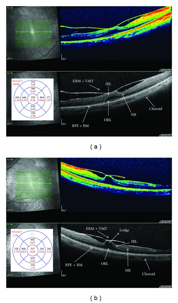
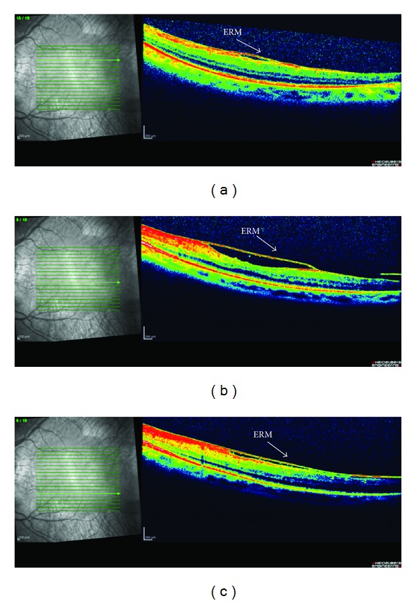
Purpose: To investigate long-term retinal changes after microincision pars plana vitrectomy surgery (MIVS) and internal limiting membrane (ILM) peeling outcome in retinitis pigmentosa (RP) patients affected by vitreomacular traction syndrome (VMT) with higher vitreous surface adhesion or coexisting epiretinal membrane (ERM).

Methods: Eight RP patients suffering from VMT were evaluated by means of best corrected visual acuity (BCVA), anterior and posterior binocular examination, spectral-domain optical coherence tomography (SD-OCT), MP-1 microperimetry (MP-1), and full-field electroretinogram (ERG), before MIVS and ILM peeling and during the 36-month follow-up. Patients were hospitalized for two days after the surgery. Surgical procedure was performed following this schedule: surgical removal of crystalline lens (MICS), MIVS with 23-gauge sutureless system trocars, core vitreous body removal, and balanced-sterile-salin-solution- (BSS-) air-gas (SF6) exchange.

Results: All patients presented visual acuity (VA) increase after MIVS. None of the patients developed ocular hypertension or vitreomacular adhesions during the 3-year follow-up. MP-1 bivariate contour ellipse area (BCEA) was reduced in its dimensions and improved in all patients demonstrating a better fixation.

Conclusions: MIVS could be the gold standard therapy in RP patients with VMT and higher vitreous surface adhesion or coexisting ERM if medical therapy is not applicable or not effective.


Figures

Figure 1
Figure 1
Spectral-domain optical coherence tomography (SD-OCT) of right (a) and left (b) eyes. Retinitis pigmentosa (RP) patients affected by vitreomacular traction syndrome (VMT) with higher vitreous surface adhesion and/or coexisting epiretinal membrane (ERM). Intraretinal macular detachment with hyporeflective and convex lodge observed in the neuroretina (NR) near the surface between inner (IRL) and outer retina (ORL) layers. Bruch's membrane (BM). Retinal pigment epithelial (RPE). Macular average thickness (μm) and volume (mm3).
Figure 2
Figure 2
Spectral-domain optical coherence tomography (SD-OCT) of left eye. Retinitis pigmentosa (RP) patient affected from vitreomacular traction syndrome (VMT) and coexisting epiretinal membrane (ERM) in area perifoveal. Scans above (a) and under the fovea (b) and (c).
Figure 3
Figure 3
Spectral-domain optical coherence tomography (SD-OCT) of right (a) and left (b) eyes. Retinitis pigmentosa (RP) patients after mini invasive vitrectomy surgery (MIVS) with internal limiting (ILM) and epiretinal membrane (ERM) peeling. Intraretinal macular detachment and the convex lodge are disappearing. The macular thickness is reduced, particularly, the outer retinal layers and retinal pigment epithelial (RPE) are thinning and impairing. Right eye (a). There are hyperreflective dots and spots in and around the external limiting membrane (ELM), inner segment/outer segment (IS/OS) junction, and RPE. Left eye (b). The length of the preserved IS/OS junction is between the limits that correspond to the two dotted lines with hyperreflective material in the outer retinal layers (ORL) causing dome-shaped elevations. Magnified view of the border where the IS/OS line disappears. The absence of the photoreceptor integrity line outside this central zone explains the patient's symptoms of very poor night vision and limited side vision. These elevations appear to correspond to pigment clumping seen on fundoscopy. The photoreceptor integrity line is present under the macula but fades outside the macula, a pattern characteristic of rod degeneration.

Similar articles

Cited by

References

    1. Heckenlively JR. RP cone-rod degeneration. Transactions of the American Ophthalmological Society. 1987;85:438–470. - PMC - PubMed
    1. Griever S, Fishman GA, Anderson RJ, Alexander KR, Derlacki DJ. Rate of visual field loss in retinitis pigmentosa. Ophthalmology. 1997;104(3):460–465. - PubMed
    1. Pruett RC. Retinitis pigmentosa: clinical observations and correlations. Transactions of the American Ophthalmological Society. 1983;81:693–735. - PMC - PubMed
    1. Berson EL. Retinitis pigmentosa: the Friedenwald lecture. Investigative Ophthalmology and Visual Science. 1993;34(5):1659–1676. - PubMed
    1. Hajali M, Fishman GA, Anderson RJ. The prevalence of cystoid macular oedema in retinitis pigmentosa patients determined by optical coherence tomography. British Journal of Ophthalmology. 2008;92(8):1065–1068. - PubMed

Publication types

LinkOut - more resources