

Understanding hydrogen sulfide storage: probing conditions for sulfide release from hydrodisulfides
- PMID: 25010540
- PMCID: PMC4120993
- DOI: 10.1021/ja505371z
Understanding hydrogen sulfide storage: probing conditions for sulfide release from hydrodisulfides
Abstract
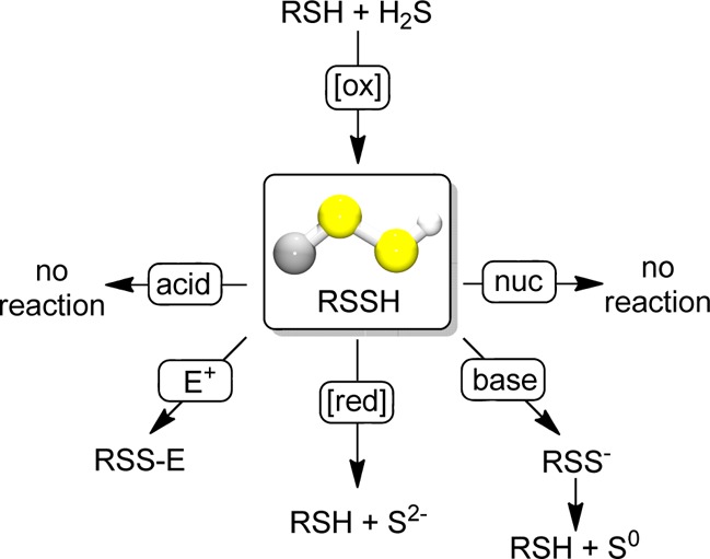
Hydrogen sulfide (H2S) is an important biological signaling agent that exerts action on numerous (patho)physiological processes. Once generated, H2S can be oxidized to generate reductant-labile sulfane sulfur pools, which include hydrodisulfides/persulfides. Despite the importance of hydrodisulfides in H2S storage and signaling, little is known about the physical properties or chemical reactivity of these compounds. We report here the synthesis, isolation, and characterization (NMR, IR, Raman, HRMS, X-ray) of a small-molecule hydrodisulfide and highlight its reactivity with reductants, nucleophiles, electrophiles, acids, and bases. Our experimental results establish that hydrodisulfides release H2S upon reduction and that deprotonation results in disproportionation to the parent thiol and S(0), thus providing a mechanism for transsulfuration in the sulfane sulfur pool.
Figures
References
-
- Wang R. Physiol. Rev. 2012, 92, 791–896. - PubMed
-
- Mustafa A. K.; Gadalla M. M.; Sen N.; Kim S.; Mu W. T.; Gazi S. K.; Barrow R. K.; Yang G. D.; Wang R.; Snyder S. H. Sci. Signal. 2009, 2, ra72. - PMC - PubMed
- Vandiver M. S.; Paul B. D.; Xu R. S.; Karuppagounder S.; Rao F.; Snowman A. M.; Ko H. S.; Il Lee Y.; Dawson V. L.; Dawson T. M.; Sen N.; Snyder S. H. Nat. Commun. 2013, 4. - PMC - PubMed
- Kessler D. FEMS Microbiol. Rev. 2006, 30, 825–840. - PubMed
- Kabil O.; Banerjee R. J. Biol. Chem. 2012, 287, 44561–44567. - PMC - PubMed
- Kimura Y.; Mikami Y.; Osumi K.; Tsugane M.; Oka J.; Kimura H. FASEB J. 2013, 27, 2451–2457. - PubMed
- Paul B. D.; Snyder S. H. Nat. Rev. Mol. Cell Biol. 2012, 13, 499–507. - PubMed
Publication types
MeSH terms
Substances
Grants and funding
LinkOut - more resources
Full Text Sources
Other Literature Sources
