Review
. 2015 Feb;21(1):88-102.
doi: 10.1089/ten.TEB.2014.0180. Epub 2014 Aug 19.

Imaging strategies for tissue engineering applications

Affiliations
Review

Imaging strategies for tissue engineering applications

Seung Yun Nam et al. Tissue Eng Part B Rev. 2015 Feb.

Abstract

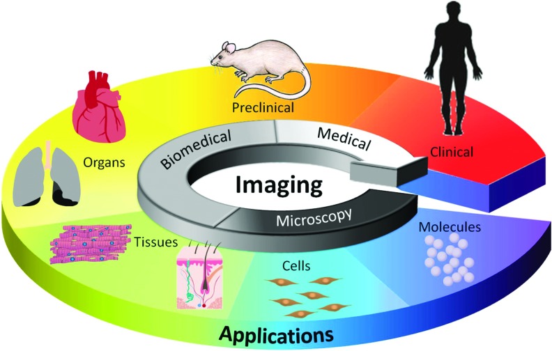
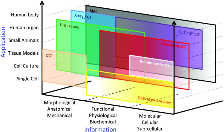
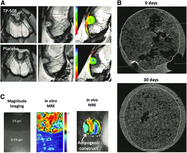
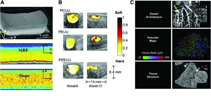
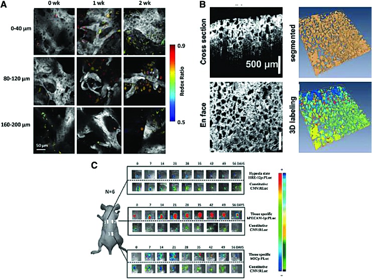
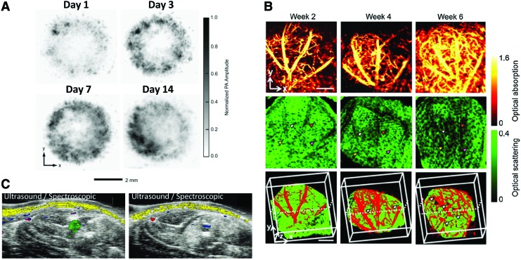
Tissue engineering has evolved with multifaceted research being conducted using advanced technologies, and it is progressing toward clinical applications. As tissue engineering technology significantly advances, it proceeds toward increasing sophistication, including nanoscale strategies for material construction and synergetic methods for combining with cells, growth factors, or other macromolecules. Therefore, to assess advanced tissue-engineered constructs, tissue engineers need versatile imaging methods capable of monitoring not only morphological but also functional and molecular information. However, there is no single imaging modality that is suitable for all tissue-engineered constructs. Each imaging method has its own range of applications and provides information based on the specific properties of the imaging technique. Therefore, according to the requirements of the tissue engineering studies, the most appropriate tool should be selected among a variety of imaging modalities. The goal of this review article is to describe available biomedical imaging methods to assess tissue engineering applications and to provide tissue engineers with criteria and insights for determining the best imaging strategies. Commonly used biomedical imaging modalities, including X-ray and computed tomography, positron emission tomography and single photon emission computed tomography, magnetic resonance imaging, ultrasound imaging, optical imaging, and emerging techniques and multimodal imaging, will be discussed, focusing on the latest trends of their applications in recent tissue engineering studies.


Figures

<b>FIG. 1.</b>
FIG. 1.
Illustration for the role of imaging in tissue engineering applications. Color images available online at www.liebertpub.com/teb
<b>FIG. 2.</b>
FIG. 2.
Available applications and information for various imaging modalities. CT, computed tomography; MRI, magnetic resonance imaging; OCT, optical coherence tomography; PET, positron emission tomography; SPECT, single photon emission computed tomography. Color images available online at www.liebertpub.com/teb
<b>FIG. 3.</b>
FIG. 3.
X-ray and CT images of various tissue-engineered scaffolds. (A) Micro-CT analysis of proteoglycan production by chondrocytes seeded in various fibroin scaffolds using Hexabrix contrast agent enhancement. Reprinted with permission from Wang et al. (B) Three-dimensional reconstructed micro-CT images of vessel ingrowth within a gel system consisting of an arteriovenous loop placed in a particulated porous hydroxyapatite and β-tricalcium phosphate matrix with and without incorporation of exogenous growth factors. Reprinted with permission from Arkudas et al. (C) Synchroton radiation CT imaging of polyurethane scaffolds seeded with endothelial cells and labeled with anti-CD34-biotin antibodies and FeO-streptavidin particles. Reprinted with permission from Thimm et al. Color images available online at www.liebertpub.com/teb
<b>FIG. 4.</b>
FIG. 4.
Nuclear medicine imaging of tissue engineering applications to evaluate wound healing. (A) Evaluation of osteogenic metabolism within a bone defect model using PET/CT imaging to assess healing following treatment with poly(lactic-co-glycolic) acid (PLGA) scaffolds seeded with bone marrow mesenchymal stem cells. Reprinted with permission from Lin et al. (B) PET/CT imaging of angiogenesis induced in a hindlimb ischemia model with αvb3 targeted dendritic nanoprobes. Reprinted with permission from van de Almutairi et al. Color images available online at www.liebertpub.com/teb
<b>FIG. 5.</b>
FIG. 5.
MR imaging to evaluate tissue-engineered scaffolds and wound healing. (A) Cartilage-sensitive MR imaging to evaluate healing within a bone defect model following delivery of thrombin peptide (TP)-508 within PLGA microspheres. Reprinted with permission from Kim et al. (B) MR imaging of SPIO-labeled chondrocytes seeded in polyvinylidene difluoride hydrogels at 0 and 30 days. Reprinted with permission from Ramaswamy et al. (C) Nondestructive monitoring of mesenchymal stem cells-based tissue-engineered constructs using high-resolution magnetic resonance elastography (MRE). Reprinted with permission from Othman et al. Color images available online at www.liebertpub.com/teb
<b>FIG. 6.</b>
FIG. 6.
Ultrasound imaging of 3D scaffolds. (A) Characterization of mineral content in collagen hydrogels using high-resolution spectral ultrasound. Reprinted with permission from Gudur et al. (B) Normalized strain maps laid over B-mode ultrasound images of various polymer scaffolds implanted in abdominal wall defects. Reprinted with permission from Yu et al. (C) Three-dimensional functional ultrasound imaging of three different liver matrix scaffolds following reseeding with hepatoblast-like cells and bioreactor cultivation to evaluate vascular architecture via contrast infusion. Reprinted with permission from Gessner et al. Color images available online at www.liebertpub.com/teb
<b>FIG. 7.</b>
FIG. 7.
Optical imaging to evaluate scaffold architecture and cell function. (A) In vitro tracking of adipose tissue structure during culture within perfusion chambers using two photon excited fluorescence imaging. Reprinted with permission from Ward et al. (B) OCT imaging to characterize EH-PEG hydrogels fabricated using different formulations in terms of volume porosity, interconnectivity, and pore size. Reprinted with permission from Chen et al. (C) In vivo monitoring of human adipose tissue-derived stromal mesenchymal cell differentiation in subcutaneous implanted demineralized bone matrix scaffolds using bioluminescence imaging. Reprinted with permission from Bago et al. Color images available online at www.liebertpub.com/teb
<b>FIG. 8.</b>
FIG. 8.
Photoacoustic imaging to track cells and monitor neovascularization. (A) In vitro photoacoustic microscopy images to evaluate the distribution of melanoma cells seeded in PLGA inverse opal scaffolds under different culture conditions. Reprinted with permission from Zhang et al. (B) Optical-resolution photoacoustic microscopy (top), OCT (middle), and combined (bottom) images of neovascularization within inverse opal scaffolds implanted in a mouse ear model. Reprinted with permission from Cai et al. (C) Combined ultrasound and spectroscopic photoacoustic imaging of gold nanoparticle-labeled mesenchymal stem cells intramuscularly injected within a PEGylated fibrin hydrogel. Reprinted with permission from Nam et al. Color images available online at www.liebertpub.com/teb

References

    1. Place E.S., Evans N.D., and Stevens M.M.Complexity in biomaterials for tissue engineering. Nat Mater 8,457, 2009 - PubMed
    1. Dvir T., Timko B.P., Kohane D.S., and Langer R.Nanotechnological strategies for engineering complex tissues. Nat Nanotechnol 6,13, 2010 - PMC - PubMed
    1. Griffith L.G., and Naughton G.Tissue engineering—current challenges and expanding opportunities. Science 295,1009, 2002 - PubMed
    1. Appel A.A., Anastasio M.A., Larson J.C., and Brey E.M.Imaging challenges in biomaterials and tissue engineering. Biomaterials 34,6615, 2013 - PMC - PubMed
    1. Cai X., Zhang Y.S., Xia Y., and Wang L.V.Photoacoustic microscopy in tissue engineering. Mater Today 16,67, 2013 - PMC - PubMed