. 2015 Feb;26(2):314-27.
doi: 10.1681/ASN.2013090985. Epub 2014 Jul 10.

GIV/girdin links vascular endothelial growth factor signaling to Akt survival signaling in podocytes independent of nephrin

Affiliations

GIV/girdin links vascular endothelial growth factor signaling to Akt survival signaling in podocytes independent of nephrin

Honghui Wang et al. J Am Soc Nephrol. 2015 Feb.

Abstract

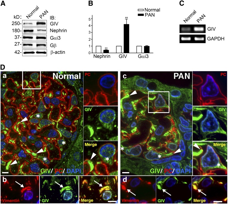
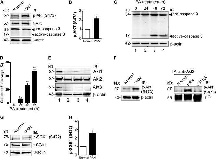
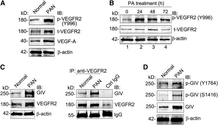
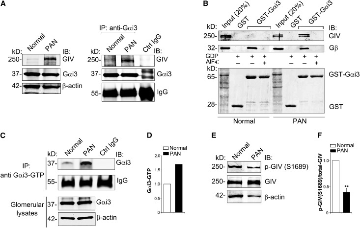
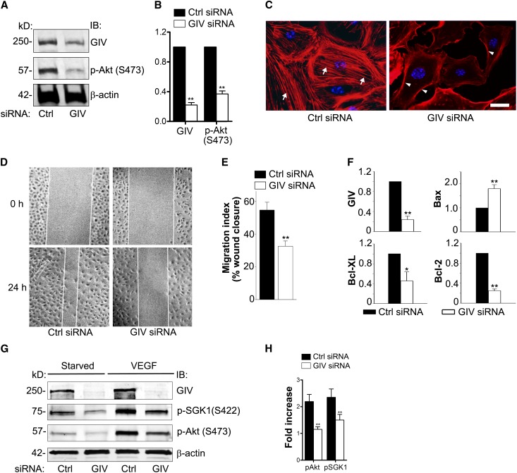
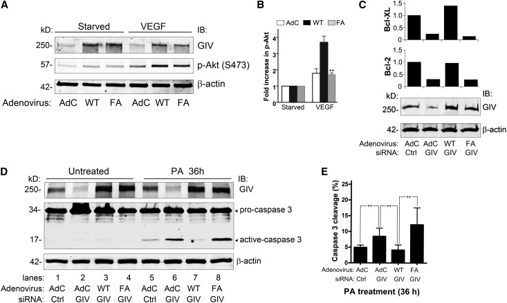
Podocytes are critically involved in the maintenance of the glomerular filtration barrier and are key targets of injury in many glomerular diseases. Chronic injury leads to progressive loss of podocytes, glomerulosclerosis, and renal failure. Thus, it is essential to maintain podocyte survival and avoid apoptosis after acute glomerular injury. In normal glomeruli, podocyte survival is mediated via nephrin-dependent Akt signaling. In several glomerular diseases, nephrin expression decreases and podocyte survival correlates with increased vascular endothelial growth factor (VEGF) signaling. How VEGF signaling contributes to podocyte survival and prevents apoptosis remains unknown. We show here that Gα-interacting, vesicle-associated protein (GIV)/girdin mediates VEGF receptor 2 (VEGFR2) signaling and compensates for nephrin loss. In puromycin aminonucleoside nephrosis (PAN), GIV expression increased, GIV was phosphorylated by VEGFR2, and p-GIV bound and activated Gαi3 and enhanced downstream Akt2, mammalian target of rapamycin complex 1 (mTORC1), and mammalian target of rapamycin complex-2 (mTORC2) signaling. In GIV-depleted podocytes, VEGF-induced Akt activation was abolished, apoptosis was triggered, and cell migration was impaired. These effects were reversed by introducing GIV but not a GIV mutant that cannot activate Gαi3. Our data indicate that after PAN injury, VEGF promotes podocyte survival by triggering assembly of an activated VEGFR2/GIV/Gαi3 signaling complex and enhancing downstream PI3K/Akt survival signaling. Because of its important role in promoting podocyte survival, GIV may represent a novel target for therapeutic intervention in the nephrotic syndrome and other proteinuric diseases.

Keywords: VEGF; cell survival; glomerular disease; podocyte.


Figures

Figure 1.
Figure 1.
GIV protein and mRNA are induced in glomeruli from PA-treated rats. (A) Expression of GIV protein is greatly increased (4-fold) and nephrin is decreased (to 40% of normal) in glomeruli from PAN rats. Gαi3 and Gβ remain unchanged. Equal aliquots (50 µg) of glomerular lysate from normal or PAN (7-day) rats are immunoblotted for GIV, nephrin, Gαi3, Gβ, and β-actin, and analyzed by Li-Cor Odyssey Infrared Imaging. (B) Quantification of data in A (normalized to β-actin). Data are expressed as the fold change in PAN compared with normal controls (**P<0.001; mean±SD n=6). (C) GIV mRNA is increased 10-fold in glomeruli after PA injury. RT-PCR is carried out on mRNA isolated from glomeruli prepared from normal and PAN rats using specific primers for rat GIV and GAPDH as a control. (D) GIV is highly expressed in podocytes in glomeruli from both normal and PAN rats. GIV is concentrated in primary processes (arrowheads) and cell bodies (asterisks) of podocytes in both normal (Da) and PAN (Dc) rats. Podocytes are identified by location and podocalyxin staining. We are unable to detect differences in the level of GIV staining between podocytes in glomeruli from normal and PAN rats. The boxed regions in A and C are enlarged (×2) to the right. Within podocytes, GIV is concentrated in primary processes (arrows) that stain for vimentin, a marker for podocytes in both normal (Db) and PAN (Dd) glomeruli. Semithin cryosections of kidney from normal and PAN-treated rats are stained for PC (red) or vimentin (red), and GIV-CT (green) and DAPI (blue), and examined by immunofluorescence. IB, immunoblot; GAPDH, glyceraldehyde-3-phosphate dehydrogenase; PC, podocalyxin; DAPI, 4,6-diamidino-2-phenylindole. Bar, 10 μm.
Figure 2.
Figure 2.
Akt/mTORC2 signaling is enhanced and podocyte survival is maintained after PA injury. (A) Phosphorylation of Akt is enhanced (2.2-fold) in glomeruli from PAN rats. Equal aliquots (50 µg) of glomerular lysate from normal or PAN (7-day) rats are immunoblotted for p-Akt (S473), t-Akt, and β-actin. (B) Quantification of data in A (normalized to β-actin). Data are expressed as the fold change in PAN compared with normal controls (**P<0.001; mean±SD n=6). (C) Podocytes survive at early stages of PA injury. At time 0, most of the caspase 3 is in the inactive (procaspase 3) form. At 24 hours, a small amount of caspase 3 cleavage (active caspase) is seen; after longer PA treatment (48 or 72 hours), the amount of cleaved (active) caspase 3 is greatly increased. In vitro differentiated mouse podocytes are treated with 30 μg/ml PA for the indicated times. Equal amounts of whole cell lysate are immunoblotted for caspase 3 and β-actin. (D) Quantification of data in C (normalized to β-actin). Data are expressed as the percentage of total caspase 3 (procaspase 3+active caspase 3) that is active (**P<0.001; mean±SD n=4). (E) Akt2 is the major Akt isoform expressed in glomeruli from rats and differentiated mouse podocytes. Equal amounts of lysate (50 μg) from C2C12 cells (positive control, lane 1), glomeruli from normal (lane 2), or PAN rats (lane 3), and differentiated mouse podocytes (lane 4) are immunoblotted for Akt1, Akt2, Akt3, and β-actin. (F) Akt2 is specifically activated in PAN. Akt2 is immunoprecipitated from glomerular lysate (1 mg) from normal or PAN rats using anti-Akt2 IgG followed by protein A magnetic beads. Bound proteins and lysates (100 μg) are immunoblotted for p-Akt (S473) and β-actin. Control IgG is incubated with a mixture of glomerular lysates from normal and PAN rats. (G) Phosphorylation of SGK1, a substrate of mTORC2 kinase, is enhanced (2.8-fold) in glomeruli from PAN rats. Equal aliquots (50 µg) of glomerular lysate from normal or PAN (7-day) rats are immunoblotted for p-SGK1(S422), t-SGK1, and β-actin. (H) Quantification of data in C (normalized to β-actin). Data are expressed as the fold change in PAN compared with normal controls (**P<0.001; mean±SD n=4). IB, immunoblot; IP, immunoprecipitate.
Figure 3.
Figure 3.
Activated VEGFR2 interacts with GIV in PAN. (A) VEGF-A is increased and phosphorylation of VEGFR2 (p-VEGFR2) is enhanced in glomeruli from PAN rats. Equal aliquots (50 μg) of glomerular lysate from normal or PAN rats are immunoblotted for p-VEGFR2 (Y996), t-VEGFR2, VEGF-A, and β-actin. (B) Activation of VEGFR2 is enhanced in podocytes after PA treatment. In vitro differentiated mouse podocytes are treated with 30 μg/ml PA for the indicated times. Equal amounts of whole cell lysate are immunoblotted for p-VEGFR2 (Y996), t-VEGFR2, and β-actin. (C) Coimmunoprecipitation of GIV with VEGFR2 is greatly increased (3.5×) in PAN glomeruli. For immunoprecipitation, glomerular lysate (1 mg) from normal or PAN rats is incubated with anti-VEGFR2 IgG followed by protein A magnetic beads. Bound proteins and lysates (100 μg) are immunoblotted for GIV, VEGFR2, and β-actin as described in the Concise Methods. Control IgG is incubated with a mixture of glomerular lysates from normal and PAN rats. (D) Phosphorylation of GIV at Y1764 and S1416 is increased in glomeruli from PAN rats. Equal aliquots (50 μg) of glomerular lysate from normal or PAN rats are immunoblotted for p-GIV (Y1764), p-GIV (S1416), GIV, and β-actin. IB, immunoblot; IP, immunoprecipitate.
Figure 4.
Figure 4.
GIV binds and activates Gαi3 upon PA injury. (A) The amount of GIV that coimmunoprecipitates with Gαi3 is greatly increased in PAN rats. Immunoprecipitation is carried out on glomerular lysates with anti-Gαi3 IgG, and bound proteins are immunoblotted for GIV, Gαi3, and β-actin as in Figure 3B. (B) GIV in glomeruli form PAN rats binds inactive (AlF4) but not active (+AlF4) Gαi3, whereas GIV from normal glomeruli does not bind either one. Gβ (positive control) binds inactive Gαi3 from both normal and PAN glomeruli. The upper panel shows GST-Gαi3 incubated with glomerular lysate (1 mg) from normal or PAN rats in the presence of GDP and AlF4 (to activate Gαi3) or GDP alone followed by immunoblotting of bound proteins for GIV and Gβ subunits. The lower panel shows Ponceau S staining. (C) Active Gαi3 (Gαi3-GTP) is increased 1.7-fold in glomeruli from PAN rats. Immunoprecipitation is carried out with an antibody that specifically recognizes only active Gαi3-GTP, and bound proteins are immunoblotted for total Gαi3 and β-actin. (D) Quantification of data in C. Data are expressed as the fold change in Gαi3-GTP level in PAN glomeruli versus normal controls. (E) Phosphorylation of GIV at S1689 is reduced in glomeruli from normal rats. Because the expression of GIV is increased 4-fold in PAN compared with normal glomeruli (Figure 1, A and B), 70 μg of normal glomerular lysate and 20 μg of PAN lysate are loaded (to equalize the total GIV) and immunoblotted for GIV, p-GIV (S1689), and β-actin. (F) Quantification of data in E (normalized to β-actin). Data are expressed as the fold change in PAN compared with normal controls (**P<0.001; mean±SD n=4). IB, immunoblot; IP, immunoprecipitate.
Figure 5.
Figure 5.
GIV binds VEGFR2 and mediates assembly of a VEGFR2/GIV/Gαi3 molecular complex in both VEGF-treated and PA-treated podocytes. (A) GIV and Gαi3 coimmunoprecipitate with VEGFR2 in VEGF-stimulated but not in serum-starved podocytes. Differentiated podocytes are serum starved or stimulated with VEGF (40 ng/ml) for 30 minutes, and cell lysates (500 μg) are incubated with anti-VEGFR2 IgG. Control IgG is incubated with a mixture of lysates from starved and VEGF-stimulated podocytes. Immunoprecipitates and cell lysates (50 μg) are immunoblotted for GIV, VEGFR2, p-Akt (S473), t-Akt, Gαi3, and β-actin. (B) Coimmunoprecipitation of both Gαi3 and GIV with VEGFR2 is greatly enhanced after PA treatment (24 hours) of differentiated podocytes, indicating that their interaction is greatly increased. Differentiated podocytes are incubated (24 hours) with or without PA (30 μg/ml), and cell lysates (500 μg) are incubated with anti-VEGFR2 or control IgG. Immunoprecipitates, and cell lysates (50 μg) are analyzed by immunoblotting with antibodies against GIV, VEGFR2, Gαi3, and β-actin. Control IgG is incubated with a mixture of podocyte lysates from untreated and PA-treated (24-hour) podocytes. IB, immunoblot; IP, immunoprecipitate.
Figure 6.
Figure 6.
GIV enhances Akt phosphorylation, actin remodeling, cell migration, and survival in podocytes. (A) p-Akt is decreased in GIV-depleted podocytes. Differentiated podocytes are transfected with control or GIV siRNA. Forty-eight hours later, cell lysates are immunoblotted for GIV, p-Akt (S473), t-Akt, and β-actin. (B) Quantification of data from 10 experiments expressed as the fold change in GIV siRNA versus Ctrl siRNA-treated podocytes (normalized to β-actin) (**P<0.001; mean±SD). (C) Podocytes treated with control siRNA show stress fibers (arrows), whereas GIV-depleted podocytes show prominent cortical actin (arrowheads). Control or GIV siRNA-treated podocytes are fixed and stained with Phalloidin Alexa Fluor 594 and DAPI. (D) Cell migration assay. Twenty-four hours after wounding, podocytes treated with control siRNA cover most of the wound area, whereas closure is impaired in GIV-depleted cells (GIV siRNA). Podocyte monolayers are scratch-wounded and examined by phase-contrast microscopy after 0 or 24 hours. (E) Quantification of data from eight experiments as in D (**P<0.001; mean±SEM). (F) After siRNA depletion of GIV (75%), proapoptotic Bax is increased (1.8-fold) and antiapoptotic Bcl-2 and Bcl-XL are decreased (to 25% and 46%, respectively) compared with controls (Ctrl siRNA). mRNA levels are determined by quantitative real-time PCR of total RNA (normalized to β-actin mRNA) (*P<0.05; **P<0.001; mean±SEM n=5). (G) VEGF-mediated phosphorylation of Akt and SGK1 is abolished in GIV-depleted podocytes. Podocytes are transfected with control or GIV siRNA by the PTD-DRBD method. Forty-eight hours after transfection, cells are serum starved or stimulated with VEGF (40 ng/ml) for 30 minutes, and cell lysates are immunoblotted for p-Akt (S473), p-SGK1 (S422), and β-actin. (H) Quantification of data from three representative experiments as in G expressed as the fold change in VEGF-mediated stimulation of p-Akt and p-SGK1 (normalized to β-actin) compared with starved controls (**P<0.001; mean±SD). DAPI, 4,6-diamidino-2-phenylindole; IB, immunoblot; Ctrl, control. Bar, 10 μm.
Figure 7.
Figure 7.
The GEF function of GIV is required for podocyte survival. (A) VEGF-mediated prosurvival Akt signaling is enhanced in podocytes overexpressing GIV-WT (WT) and is inhibited in podocytes overexpressing GIV-FA (FA). Podocytes are transduced with Ad-GIV-WT (WT) or Ad-GIV-FA (FA), or control adenovirus (AdC). Forty-eight hours later, they are switched to 0.4% serum for 24 hours and then stimulated with VEGF (40 ng/ml) for 30 minutes. Equal amounts of cell lysate are immunoblotted for GIV, p-Akt (S473), and β-actin. (B) Quantification of data in A (normalized to β-actin). Data are expressed as the fold change in AdC–, Ad-GIV-WT–, or Ad-GIV-FA–treated podocytes. (C) Depletion of endogenous GIV in cells transduced with control virus (AdC) decreases expression of the antiapoptotic markers Bcl-2 (30%) and Bcl-XL (25%). Overexpression of GIV-WT prevents this reduction in expression of Bcl-2 and Bcl-XL, whereas overexpression of GIV-FA does not. Podocytes are transduced with control (AdC), Ad-GIV-WT, or Ad-GIV-FA, followed (24 hours later) by control or GIV siRNA. After 48 hours, equal amounts of cell lysates are immunoblotted for GIV or β-actin. RNA is isolated and relative mRNA levels of Bcl-2 and Bcl-XL are determined by quantitative real-time PCR. (D) Addition of PA (36 hours) to GIV siRNA-treated podocytes transduced with control virus (lane 6) leads to increased caspase 3 cleavage (1.7-fold) compared with control siRNA-treated cells (lane 5). Overexpression GIV-WT (lane 7) inhibits the PA-mediated increase in caspase 3 cleavage but overexpression of GIV-FA (lane 8) does not. No caspase cleavage is seen in untreated cells (lanes 1–4). Podocytes are transduced with control virus (AdC), Ad-GIV-WT, or Ad-GIV-FA, followed (24 hours later) by control or GIV siRNA. After 48 hours, they are treated with 30 µg/ml PA for 36 hours. Equal amounts of cell lysates are immunoblotted for GIV, caspase 3, and β-actin. (E) Quantification of data in D (normalized to β-actin). Data are expressed as the percentage of active caspase 3/total caspase 3 (procaspase 3+active caspase 3) (**P<0.001; mean±SD n=3). IB, immunoblotting.
Figure 8.
Figure 8.
Working model of GIV’s role in early podocyte injury in PAN nephrosis. (A) In normal podocytes, Akt survival signaling is mediated through the well defined nephrin-dependent PI3K/Akt pathway. (B) At early time points (7 days) after PAN injury, increased GIV expression coincides with decreased nephrin expression and activation of VEGFR2 and Akt. GIV binds to activated VEGFR2 and is tyrosine phosphorylated by VEGFR2. Activated GIV binds and activates Gαi3 (Gαi3-GTP). A VEGFR2/GIV/Gαi3 signaling complex is assembled and prolongs podocyte survival in PAN via activation of PI3K, Akt, mTORC1, and mTORC2. Thus, after PA injury when nephrin levels decrease, GIV takes over the task of enhancing Akt/mTOR survival signaling in response to VEGF.

References

    1. Greka A, Mundel P: Cell biology and pathology of podocytes. Annu Rev Physiol 74: 299–323, 2012 - PMC - PubMed
    1. Pavenstädt H, Kriz W, Kretzler M: Cell biology of the glomerular podocyte. Physiol Rev 83: 253–307, 2003 - PubMed
    1. Garg P, Holzman LB: Podocytes: Gaining a foothold. Exp Cell Res 318: 955–963, 2012 - PubMed
    1. Kerjaschki D: Caught flat-footed: Podocyte damage and the molecular bases of focal glomerulosclerosis. J Clin Invest 108: 1583–1587, 2001 - PMC - PubMed
    1. Reiser J, Sever S: Podocyte biology and pathogenesis of kidney disease. Annu Rev Med 64: 357–366, 2013 - PMC - PubMed

Publication types

MeSH terms

Substances

LinkOut - more resources