

Comparative RNA-sequencing analysis of myocardial and circulating small RNAs in human heart failure and their utility as biomarkers
- PMID: 25012294
- PMCID: PMC4121804
- DOI: 10.1073/pnas.1401724111
Comparative RNA-sequencing analysis of myocardial and circulating small RNAs in human heart failure and their utility as biomarkers
Abstract
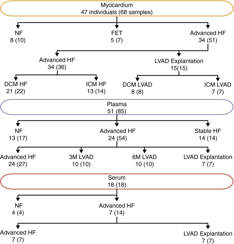
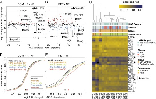
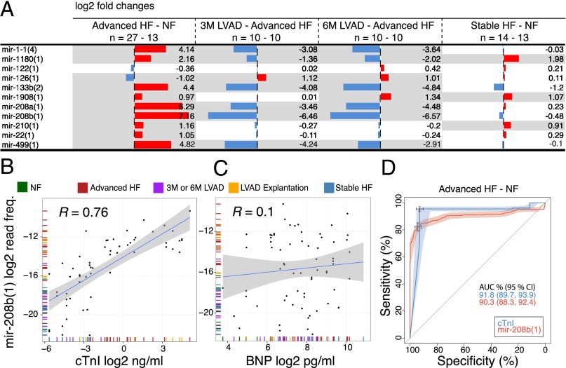
Heart failure (HF) is associated with high morbidity and mortality and its incidence is increasing worldwide. MicroRNAs (miRNAs) are potential markers and targets for diagnostic and therapeutic applications, respectively. We determined myocardial and circulating miRNA abundance and its changes in patients with stable and end-stage HF before and at different time points after mechanical unloading by a left ventricular assist device (LVAD) by small RNA sequencing. miRNA changes in failing heart tissues partially resembled that of fetal myocardium. Consistent with prototypical miRNA-target-mRNA interactions, target mRNA levels were negatively correlated with changes in abundance for highly expressed miRNAs in HF and fetal hearts. The circulating small RNA profile was dominated by miRNAs, and fragments of tRNAs and small cytoplasmic RNAs. Heart- and muscle-specific circulating miRNAs (myomirs) increased up to 140-fold in advanced HF, which coincided with a similar increase in cardiac troponin I (cTnI) protein, the established marker for heart injury. These extracellular changes nearly completely reversed 3 mo following initiation of LVAD support. In stable HF, circulating miRNAs showed less than fivefold differences compared with normal, and myomir and cTnI levels were only captured near the detection limit. These findings provide the underpinning for miRNA-based therapies and emphasize the usefulness of circulating miRNAs as biomarkers for heart injury performing similar to established diagnostic protein biomarkers.
Keywords: body fluids; cardiovascular disease; development; exRNA; miRNA-mRNA regulation.
Conflict of interest statement
Conflict of interest statement: T.T. is a cofounder of and scientific advisor to Alnylam Pharmaceuticals and a scientific advisor to Regulus Therapeutics.
Figures

References
-
- Weiland M, Gao XH, Zhou L, Mi QS. Small RNAs have a large impact: Circulating microRNAs as biomarkers for human diseases. RNA Biol. 2012;9(6):850–859. - PubMed
-
- Braun JE, Huntzinger E, Izaurralde E. The role of GW182 proteins in miRNA-mediated gene silencing. Adv Exp Med Biol. 2013;768:147–163. - PubMed
-
- Valadi H, et al. Exosome-mediated transfer of mRNAs and microRNAs is a novel mechanism of genetic exchange between cells. Nat Cell Biol. 2007;9(6):654–659. - PubMed
-
- Laterza OF, et al. Plasma microRNAs as sensitive and specific biomarkers of tissue injury. Clin Chem. 2009;55(11):1977–1983. - PubMed
Publication types
MeSH terms
Substances
Associated data
- Actions
Grants and funding
- UL1 TR000043/TR/NCATS NIH HHS/United States
- HL073029/HL/NHLBI NIH HHS/United States
- R00 HL109133/HL/NHLBI NIH HHS/United States
- P30HL101272/HL/NHLBI NIH HHS/United States
- K23HL095742/HL/NHLBI NIH HHS/United States
- UL1 TR001073/TR/NCATS NIH HHS/United States
- 8UL1TR000043/TR/NCATS NIH HHS/United States
- HD068546/HD/NICHD NIH HHS/United States
- R01 HL073029/HL/NHLBI NIH HHS/United States
- K99 HL109133/HL/NHLBI NIH HHS/United States
- K23 HL095742/HL/NHLBI NIH HHS/United States
- K08 HD068546/HD/NICHD NIH HHS/United States
- P30 HL101272/HL/NHLBI NIH HHS/United States
- HHMI/Howard Hughes Medical Institute/United States
- UL1 RR024156/RR/NCRR NIH HHS/United States
- U19CA179564/CA/NCI NIH HHS/United States
- U19 CA179564/CA/NCI NIH HHS/United States
- UL1RR024156/RR/NCRR NIH HHS/United States
- T32 GM007739/GM/NIGMS NIH HHS/United States
LinkOut - more resources
Full Text Sources
Other Literature Sources
Medical
Molecular Biology Databases
Research Materials
Miscellaneous
