. 2014 Jul 11;345(6193):220-2.
doi: 10.1126/science.1254522.

Bacterial cell wall. MurJ is the flippase of lipid-linked precursors for peptidoglycan biogenesis

Affiliations

Bacterial cell wall. MurJ is the flippase of lipid-linked precursors for peptidoglycan biogenesis

Lok-To Sham et al. Science. .

Abstract

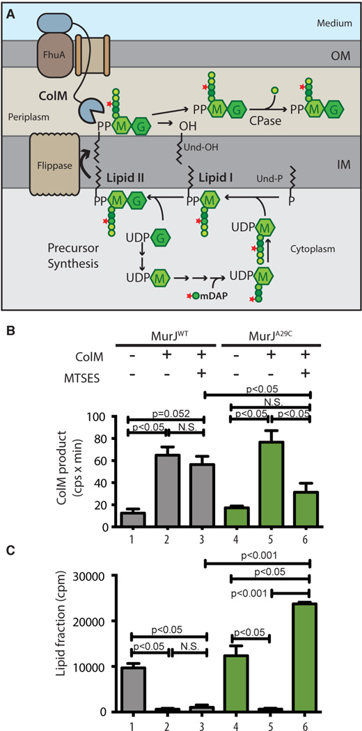
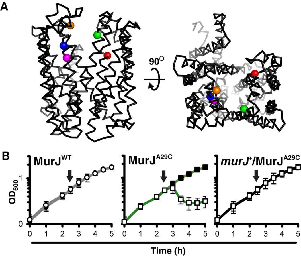
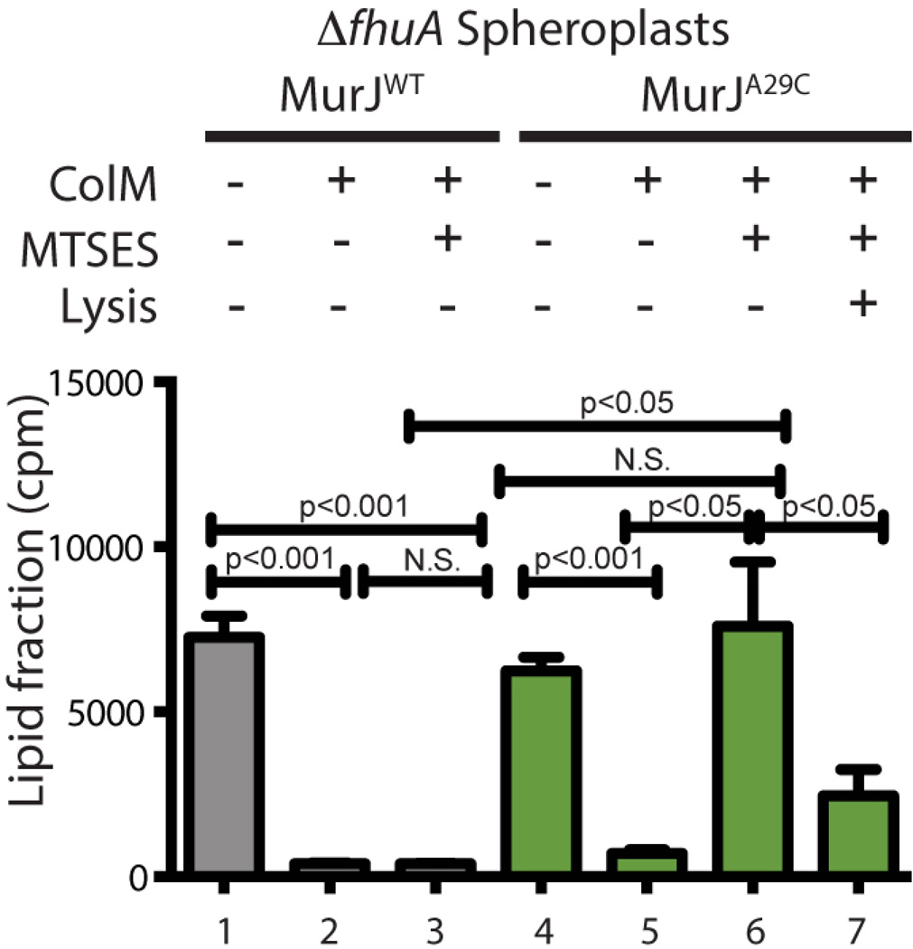
Peptidoglycan (PG) is a polysaccharide matrix that protects bacteria from osmotic lysis. Inhibition of its biogenesis is a proven strategy for killing bacteria with antibiotics. The assembly of PG requires disaccharide-pentapeptide building blocks attached to a polyisoprene lipid carrier called lipid II. Although the stages of lipid II synthesis are known, the identity of the essential flippase that translocates it across the cytoplasmic membrane for PG polymerization is unclear. We developed an assay for lipid II flippase activity and used a chemical genetic strategy to rapidly and specifically block flippase function. We combined these approaches to demonstrate that MurJ is the lipid II flippase in Escherichia coli.


Figures

Fig. 1
Fig. 1. In vivo assay for lipid II flippase activity
(A) PG precursor synthesis starts with the conversion of UDP-N-acetylglucosamine (UDP-G) to UDP-N-acetylmuramic acid (UDP-M), followed by the addition of amino acids (represented by colored spheres) to UDP-M to form the pentapeptide (pep5) stem (L-Ala-γ-D-Glu-m-DAP-D-Ala-D-Ala). [3H]-mDAP label is indicated by the star. The UDP-sugars are transferred to undecaprenol-P (Und-P) in the IM to form lipid II, which is flipped across the IM to expose the disaccharide-pep5 (Mpep5-G) for polymerization and crosslinking into PG (not illustrated). Exogenous ColM binds to FhuA and is translocated across the OM presumably through a porin. In the periplasm, ColM cleaves lipid II into undecaprenol (Und-OH) and soluble PP-Mpep5-G, which is further processed by carboxypeptidases (CPase) to produce PP-Mpep4-G. (B-C) Cells of ΔmurJ ΔlysA strains producing FLAGMurJ lacking endogenous Cys residues referred to as MurJWT (NR2592) or its derivative MurJA29C (NR2593) were labeled with [3H]-mDAP. After 15 min, ColM and/or MTSES were added as indicated and growth was continued for 10 min. Samples were then withdrawn and either extracted with hot water alone or sequentially with water then butanol. Hot water extracts were subjected to high-performance liquid chromatography (HPLC) and radiodetection to quantify the labeled ColM product (B); scintillation counting was used to quantify label in the lipid (butanol) fraction (C). See figs. S1–S4 for experimental details and peak identification. Shown are the mean ± SD; N = 3. P value determined with Student’s t test. N.S., not significant.
Fig. 2
Fig. 2. MTSES specifically inhibits the function of MurJA29C
(A) Structural model of MurJ (8). Sensitivity to MTSES is limited to specific residues within the MurJ cavity: residues 29 (green) in transmembrane domain (TMD) 1, 49 (red) in TMD 2 , and 263 (orange), 269 (blue) and 273 (magenta) in TMD 8. (B) Effect of MTSES on the growth of haploid cells producing MurJWT (left) or MurJA29C in glucose M63 medium. Lysis of MurJA29C cells is suppressed by the presence of a wild-type murJ allele (right). Arrows indicate time of MTSES addition; filled marker, no MTSES; empty marker, MTSES treated. Data represent mean ± SD; N = 3. See fig. S5 for MTSES sensitivity of other variants.
Fig. 3
Fig. 3. MurJ activity is required for ColM-dependent cleavage of lipid II in spheroplasts
Cells lacking the ColM receptor FhuA and producing the indicated MurJ variants were grown, labeled, and treated with MTSES as for figure 1. Spheroplasts were then prepared. In all but one case, spheroplasts were pelleted and resuspended in ColM reaction buffer with sucrose and added MTSES (0.8 mM) as indicated. The Lysis + sample was resuspended in buffer lacking sucrose to lyse the spheroplasts. ColM (100 µg) was added to the prepared spheroplasts as indicated and they were incubated for 15 min at 37°C. Lipid intermediates were detected after butanol extraction by scintillation counting. Statistics are as for figure 1.

Comment in

References

    1. Typas A, Banzhaf M, Gross CA, Vollmer W. From the regulation of peptidoglycan synthesis to bacterial growth and morphology. Nat Rev Microbiol. 2012 Feb;10:123. - PMC - PubMed
    1. Bouhss A, Trunkfield AE, Bugg TD, Mengin-Lecreulx D. The biosynthesis of peptidoglycan lipid-linked intermediates. FEMS Microbiol Rev. 2008 Mar;32:208. - PubMed
    1. de Pedro MA, Donachie WD, Holtje JV, Schwarz H. Constitutive septal murein synthesis in Escherichia coli with impaired activity of the morphogenetic proteins RodA and penicillin-binding protein 2. J Bacteriol. 2001 Jul;183:4115. - PMC - PubMed
    1. Inoue A, et al. Involvement of an essential gene mviN, in murein synthesis in Escherichia coli. J Bacteriol. 2008 Nov;190:7298. - PMC - PubMed
    1. Khattar MM, Begg KJ, Donachie WD. Identification of FtsW and characterization of a new ftsW division mutant of Escherichia coli. J Bacteriol. 1994 Dec;176:7140. - PMC - PubMed

Publication types

MeSH terms

Substances