. 2014 Aug;42(14):9447-60.
doi: 10.1093/nar/gku578. Epub 2014 Jul 10.

Phospho-dependent and phospho-independent interactions of the helicase UPF1 with the NMD factors SMG5-SMG7 and SMG6

Affiliations

Phospho-dependent and phospho-independent interactions of the helicase UPF1 with the NMD factors SMG5-SMG7 and SMG6

Sutapa Chakrabarti et al. Nucleic Acids Res. 2014 Aug.

Abstract

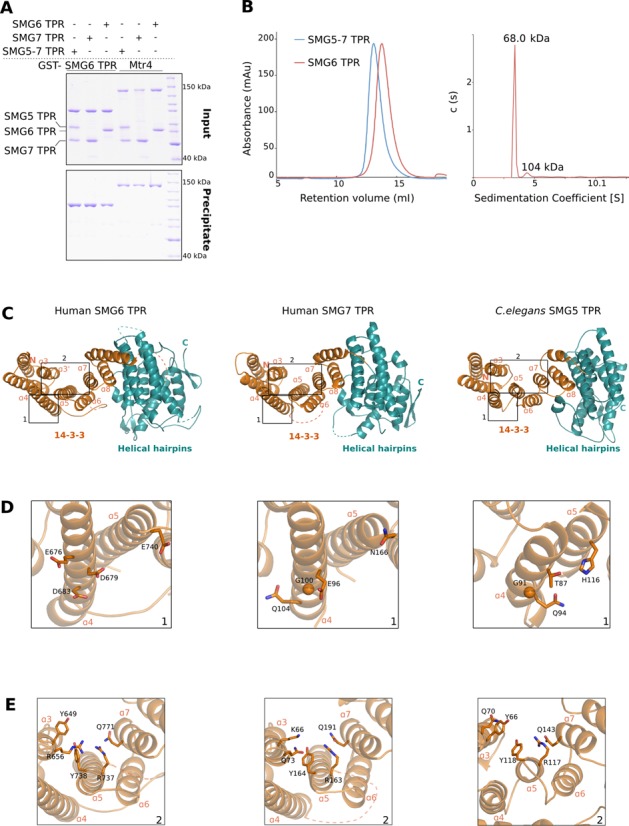
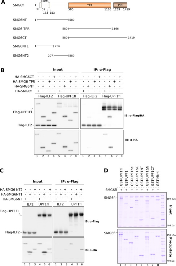
Nonsense-mediated mRNA decay (NMD) is a eukaryotic surveillance pathway that recognizes mRNAs with premature stop codons and targets them for rapid degradation. Evidence from previous studies has converged on UPF1 as the central NMD factor. In human cells, the SMG1 kinase phosphorylates UPF1 at the N-terminal and C-terminal tails, in turn allowing the recruitment of the NMD factors SMG5, SMG6 and SMG7. To understand the molecular mechanisms, we recapitulated these steps of NMD in vitro using purified components. We find that a short C-terminal segment of phosphorylated UPF1 containing the last two Ser-Gln motifs is recognized by the heterodimer of SMG5 and SMG7 14-3-3-like proteins. In contrast, the SMG6 14-3-3-like domain is a monomer. The crystal structure indicates that the phosphoserine binding site of the SMG6 14-3-3-like domain is similar to that of SMG5 and can mediate a weak phospho-dependent interaction with UPF1. The dominant SMG6-UPF1 interaction is mediated by a low-complexity region bordering the 14-3-3-like domain of SMG6 and by the helicase domain and C-terminal tail of UPF1. This interaction is phosphorylation independent. Our study demonstrates that SMG5-SMG7 and SMG6 exhibit different and non-overlapping modes of UPF1 recognition, thus pointing at distinguished roles in integrating the complex NMD interaction network.


Figures

Figure 1.
Figure 1.
Interaction of human SMG5, SMG6 and SMG7 with in vitro phosphorylated UPF1. (A) Schematic representation of the domain organization of full-length (fl) human UPF1, SMG5, SMG6 and SMG7. Folded domains are shown as rectangles and low-complexity sequences as lines. In UPF1, the CH domain (in green) and the ATPase core domains (RecA1 and 2, 1B and 1C, colored yellow, orange and red, respectively) are indicated. The regions N- and C-terminal of the core contain the SMG1 phosphorylation sites. The TPR domains of SMG5, SMG6 and SMG7 comprise the 14–3–3 and helical hairpins domain, which are colored orange and teal, respectively. The SMG5 TPR domain is bifurcated by a long linker, which was deleted in the construct used in this study. The N-terminal EBMs of SMG6 are shown as boxes. In brown are the active and catalytically dead (d) PIN domains at the C-terminus of SMG6 and SMG5, respectively. (B) Left panel: SDS-PAGE analysis of catalytically active (wt) and inactive (kinase-dead, KD) SMG1 proteins purified from HEK 293T cells. The asterisk (*) indicates a contaminant (methylosome) that co-purifies with SMG1. Right panel: in vitro kinase assay performed using purified SMG1 proteins and UPF1fl as a substrate. A corresponding Coomassie-stained gel of the radioactive kinase assay (using γ-32P ATP) indicates the enzyme: substrate ratio employed in the assay. (C) GST pull-down assays of GST-UPF1fl (treated with active SMG1 wt or inactive SMG1 KD mutant) and SMG5–7 TPR, SMG7 TPR, SMG6 TPR and SMG6fl. GST-Mtr4 was used as a negative control in this and all other GST pull-down experiments. One-fifth of the reaction mixture was used as input. Inputs and the bound fractions (precipitates) were analyzed on 8% SDS-PAGE gels and are shown in the top and bottom panels, respectively. The SMG5–7 TPR complex showed a significant affinity toward phospho-UPF1fl while SMG6fl binds UPF1fl in a phosphorylation-independent manner. The TPR domains of SMG6 and SMG7 exhibit only a weak affinity toward UPF1fl.
Figure 2.
Figure 2.
The SMG5–7 TPR complex binds the extreme C-terminal segment of UPF1. (A) Top panel: schematic representation of the UPF1 constructs designed to map the binding site of SMG5–7 TPR on UPF1. Bottom panel: GST pull-down assays of SMG5–7 TPR with the GST-UPF1 constructs described in the top panel. The assays were carried out in the presence of either SMG1 wt or SMG1 KD mutant as described for Figure 1C. The inputs and precipitates were analyzed on 10% SDS-PAGE gels. In the presence of SMG1 wt, the 200-residue stretch following the UPF1 helicase domain is sufficient for binding SMG5–7 TPR (see also Supplementary Figure S1B). (B) Top panel: schematic representation of the GST-UPF1 C-terminal constructs designed to map the SQ motifs that mediate binding of SMG5–7 TPR to UPF1. Bottom panel: GST pull-down assays (performed as described above) suggest that residues S1096 and S1116, when phosphorylated, act as interaction motifs for SMG5–7 TPR (see also Supplementary Figure S1C). The inputs and precipitates were analyzed on 15% SDS-PAGE gels. The asterisk (*) indicates a degradation product of GST-UPF1CT.
Figure 3.
Figure 3.
Overall structure of the SMG6 TPR domain. (A) GST pull-down assays of GST-SMG6 TPR with that of other TPR domains, carried out as in Figure 1C. The samples were analyzed on 10% SDS-PAGE gels; panels for inputs and precipitates are indicated. While the TPR domains of SMG5 and SMG7 interact to form a stable complex, that of SMG6 is unable to mediate such interactions. (B) Left panel: size-exclusion analyses of SMG5–7 TPR and SMG6 TPR proteins. The purified SMG5–7 TPR complex and SMG6 TPR were separately injected on a semi-analytical gel-filtration column (Superdex 200 HR 10/30, GE Healthcare) and the chromatograms were overlaid. The size-exclusion analyses demonstrate that SMG6 TPR is a monomer in solution (molecular weight of 68 kDa) in contrast to the dimeric SMG5–7 TPR (molecular weight of 123 kDa). Right panel: sedimentation velocity AUC of SMG6 TPR. SMG6 TPR was monitored at 280 nm and 50 000 rpm at 20°C for 16 h. The molecular weight of SMG6 was estimated from the sedimentation coefficient and the diffusion coefficient. (C) Overall crystal structure of the TPR domains of SMG6 (left), human SMG7 (middle, PDB ID 1YAO) and C. elegans SMG5 (right, PDB ID 3ZHE). The structures are shown in the same orientation after optimal superposition of their N-terminal 14–3–3 domain. The helices within the N-terminal 14–3–3 domain (colored orange) encompass the phosphoserine-binding motif while the helices of the C-terminal domain (colored teal) are arranged into helical hairpins. The highlighted region 1 depicts the hotspot of dimerization in the 14–3–3 domains, as derived from the structure of C. elegans SMG5–7 TPR. The highlighted region 2 indicates the phosphoserine-binding pocket within the 14–3–3 domains of the three proteins. This and all other structure figures were generated using PyMOL (http://www.pymol.org). (D) A close-up view of the highlighted region 1 from Figure 3C. A conserved glycine residue in helix α4 of SMG7 and SMG5 (middle and right panels) is at the center of the SMG5–7 TPR interface. The equivalent residue in helix α4 of SMG6 (left panel) is an asparate (D683, see also Supplementary Figure S3). (E) A close-up view of the highlighted region 2 from Figure 3C. The residues lining the phosphoserine-binding pockets of SMG6, SMG7 and SMG5 are highlighted.
Figure 4.
Figure 4.
The N-terminal domain of SMG6 encompasses multiple discrete protein–protein interaction motifs. (A) Schematic representation of the SMG6 constructs designed to map the binding site of UPF1 on SMG6. (B) co-IP assays of Flag-UPF1fl and the indicated HA-SMG6 constructs (SMG6fl, SMG6NT, SMG6 TPR and SMG6CT). SMG6 constructs were co-transfected with UPF1 in HEK-293T cells. Cell lysates were subjected to IP using an anti-Flag antibody. 2% of the total cell lysate of every sample was used as the input. Inputs and precipitates were analyzed by SDS-PAGE and immuno blotting using the indicated antibodies and are depicted on the left and right panels, respectively. Flag-ILF2 was used as a negative control in this and other co-IP experiments, which were carried out in the absence of RNase A. Only the N-terminal domain of SMG6 was precipitated by Flag-UPF1 in a manner similar to SMG6fl, indicating the presence of a UPF1 interaction motif within this region. (C) Co-IP assays of Flag-UPF1fl and HA-tagged SMG6 N-terminal constructs (SMG6NT, SMG6NT1 and SMG6NT2). The experiment was performed as described above. The phospho-independent UPF1 interaction motif of SMG6 localized to a stretch that is proximal to the TPR domain and distinct from the EBMs located within the first 160 residues. (D) GST pull-down assays of different GST-UPF1 constructs (described in Figure 2A) and SMG6fl. The pull-downs and analysis were carried out as described in Figure 1C. The inputs and precipitates are shown on the top and bottom panels, respectively. Both the CH-helicase core and the C-terminal tail of UPF1 are important for SMG6 recognition.
Figure 5.
Figure 5.
Assembly of complexes of multiple NMD factors in the presence of SMG6. (A) GST pull-down assays of GST-UPF1fl and SMG6fl in the presence of the NMD factors UPF2, UPF3 and the EJC. The pull-down was performed as described in Figure 1C. The inputs and precipitates were analyzed on 4–12% Bis-Tris gels (Invitrogen) and are depicted in the top and bottom panels, respectively. SMG6fl simultaneously interacted with UPF1 (even in the presence of UPF2 and UPF3) and the EJC, using its two distinct interaction motifs. The assembly of a large NMD complex consisting of SMG6, UPF1, UPF2, UPF3 and EJC is shown in lane 7 (see also Supplementary Figure S5). (B) GST pull-down assays of GST-UPF1fl (treated with active SMG1 wt or inactive SMG1 KD mutant) and SMG6fl and SMG5–7 TPR. The pull-downs and analysis were carried out as in Figure 1C. Top and bottom panels depict inputs and precipitates, respectively. Upon phosphorylation, GST-UPF1 simultaneously interacted with SMG6fl and SMG5–7 TPR, indicating that binding of the different TPR proteins to the C-terminus of UPF1 can occur concomitantly. (C) Model depicting the possible transient SMG6–UPF–EJC complexes assembled in the course of NMD, recapitulating the results from this study and previous reports. UPF1 (colored yellow) is depicted as a bilobal structure, with the small circle representing the N-terminal CH domain and the large circle representing the ATPase core. N-and C-terminal extensions of UPF1 are shown as yellow lines and phosphorylation sites therein are depicted as red open circles. UPF2 (in light green) binds the CH domain of UPF1 and UPF3 (colored dark green), forming the UPF complex. The TPR domains of SMG5 and SMG7 (colored purple and magenta) bind the C-terminal phospho sites of UPF1, while SMG6 TPR (colored orange) is thought to bind the N-terminal phospho site of UPF1. The N-terminus of SMG6 mediates a strong interaction with UPF1 and is crucial for its recruitment to the UPF–EJC complex. The interaction between UPF1 and the EJC (colored blue) can either be bridged by the EBM of UPF3 (left panel) or by the EBMs of SMG6 (right panel).

References

    1. Lykke-Andersen J., Bennett E.J. Protecting the proteome: eukaryotic cotranslational quality control pathways. J. Cell Biol. 2014;204:467–476. - PMC - PubMed
    1. Shoemaker C.J., Green R. Translation drives mRNA quality control. Nat. Struct. Mol. Biol. 2012;19:594–601. - PMC - PubMed
    1. Bhuvanagiri M., Schlitter A.M., Hentze M.W., Kulozik A.E. NMD: RNA biology meets human genetic medicine. Biochem. J. 2010;430:365–377. - PubMed
    1. Kervestin S., Jacobson A. NMD: a multifaceted response to premature translational termination. Nat. Rev. Mol. Cell Biol. 2012;13:700–712. - PMC - PubMed
    1. Popp M.W., Maquat L.E. Organizing principles of mammalian nonsense-mediated mRNA decay. Ann. Rev. Genet. 2013;47:139–165. - PMC - PubMed

Publication types

Associated data