. 2014 Jul;42(13):8648-62.
doi: 10.1093/nar/gku579. Epub 2014 Jul 10.

Phosphorothioate oligonucleotides can displace NEAT1 RNA and form nuclear paraspeckle-like structures

Affiliations

Phosphorothioate oligonucleotides can displace NEAT1 RNA and form nuclear paraspeckle-like structures

Wen Shen et al. Nucleic Acids Res. 2014 Jul.

Abstract

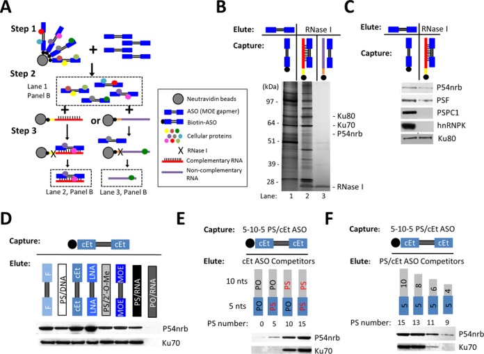
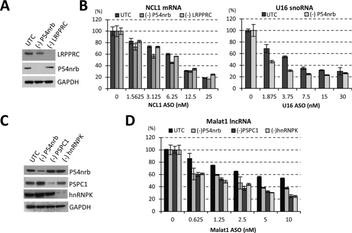
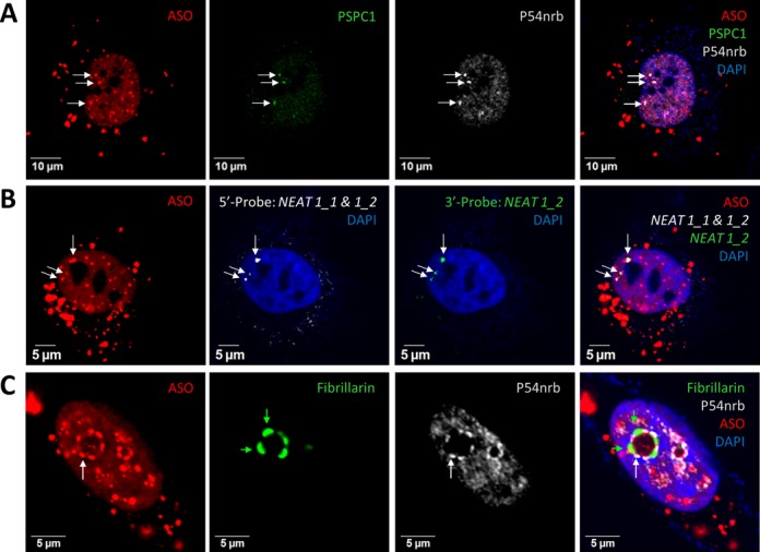
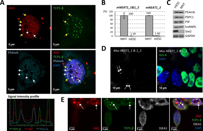
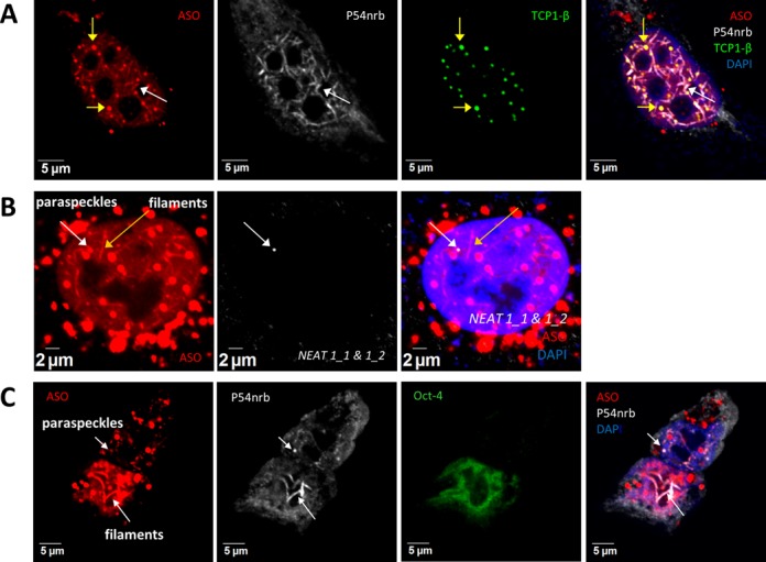
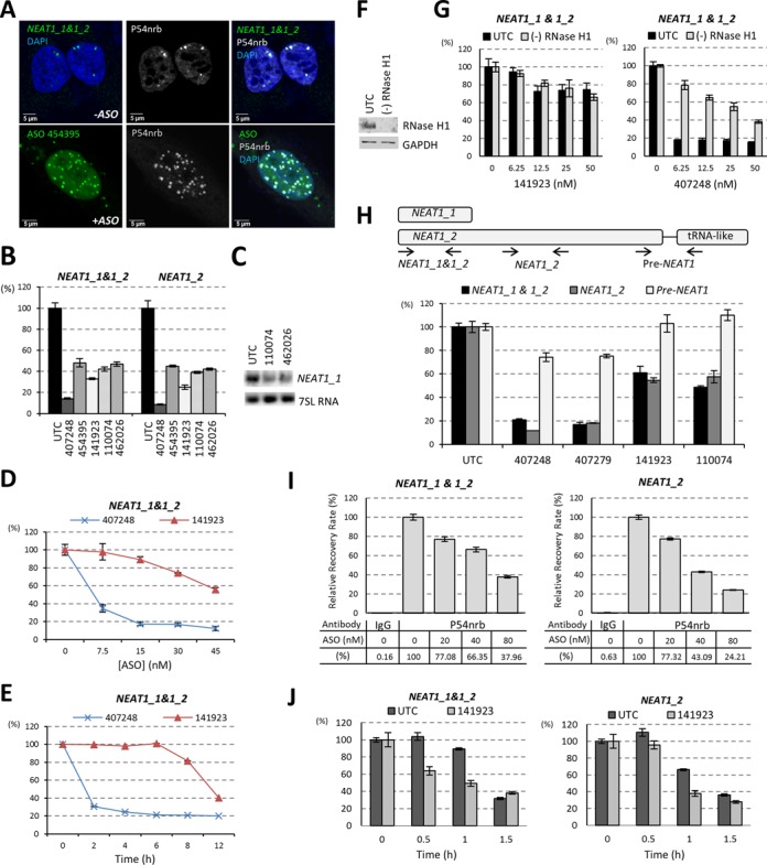
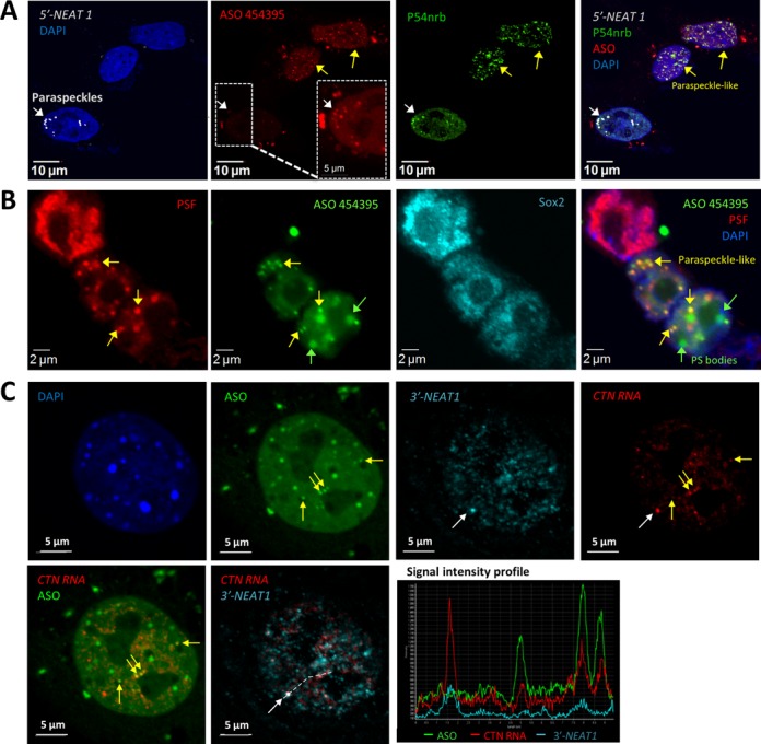
Nuclear paraspeckles are built co-transcriptionally around a long non-coding RNA, NEAT1. Here we report that transfected 20-mer phosphorothioate-modified (PS) antisense oligonucleotides (ASOs) can recruit paraspeckle proteins to form morphologically normal and apparently functional paraspeckle-like structures containing no NEAT1 RNA. PS-ASOs can associate with paraspeckle proteins, including P54nrb, PSF, PSPC1 and hnRNPK. NEAT1 RNA can be displaced by transfected PS-ASO from paraspeckles and rapidly degraded. Co-localization of PS-ASOs with P54nrb was observed in canonical NEAT1-containing paraspeckles, in perinucleolar caps upon transcriptional inhibition, and importantly, in paraspeckle-like or filament structures lacking NEAT1 RNA. The induced formation of paraspeckle-like and filament structures occurred in mouse embryonic stem cells expressing little or no NEAT1 RNA, suggesting that PS-ASOs can serve as seeding molecules to assemble paraspeckle-like foci in the absence of NEAT1 RNA. Moreover, CTN, an RNA reported to be functionally retained in paraspeckles, was also observed to localize to paraspeckle-like structures, implying that paraspeckle-like structures assembled on PS-ASOs are functional. Together, our results indicate that functional paraspeckles can form with short nucleic acids other than NEAT1 RNA.


Figures

Figure 1.
Figure 1.
Paraspeckle proteins associate with PS-ASOs. (A) Schematic representation of experimental procedures of affinity selection of proteins bound to PS-ASO/RNA duplex. (B) Silver staining of co-selected proteins with ssPS-ASO (lane 1), PS-ASO/RNA duplex (lane 2) or control ssRNA (lane 3). (C) Western analysis followed by ASO pull-down confirmed the association of paraspeckle proteins with PS-ASO or PS-ASO/RNA duplex. (D) 2′-modifications of PS-ASOs can influence protein binding. The proteins captured with 5–10–5 cEt PS-ASO were eluted by competition with 5–10–5 ASOs bearing the same sequence but with distinct 2′-modifications, or with uniform ASOs with or without PS backbone modification, as indicated above lanes. The levels of P54nrb and Ku70 were determined by western analysis. (E and F) P54nrb/ASO interaction requires 10 or more PS backbone modified nts. ASO-associated proteins captured with 5–10–5 cEt PS-ASO were eluted with 15-mer ASOs containing different numbers of PS modified nts (panel E), or with PS-ASOs with varying length (panel F), and subjected to western analysis.
Figure 2.
Figure 2.
Reduction of paraspeckle proteins increases the activity of RNase H1-based ASOs. (A) siRNA-mediated reduction of P54nrb and LRPPRC proteins, as determined by western analysis. UTC, mock treated control cells. GAPDH served as a loading control. (B) Reduction of P54nrb, but not LRPPRC, increased the activity of ASO-mediated cleavage of NCL1 mRNA (left panel) or U16 snoRNA (right panel), as determined by qRT-PCR analysis. (C) siRNA-mediated reduction of paraspeckle proteins P54nrb, PSPC1 and hnRNPK, as determined by western analysis. GAPDH served as a loading control. (D) Reduction of other paraspeckle proteins can also increase ASO activity, as exemplified with an ASO targeting Malat1 lncRNA, as determined by qRT-PCR. The error bars represent standard deviation from three parallel experiments.
Figure 3.
Figure 3.
PS-ASOs can localize to bona fide paraspeckles. (A) Co-localization of transfected PS-ASOs with paraspeckle marker proteins PSPC1 and P54nrb in HeLa cells, as indicated by arrows. The nucleus was stained with DAPI. (B) Co-localization of PS-ASOs with structural RNA NEAT1 in paraspeckles, as indicated by arrows. NEAT1 RNA was detected by RNA-FISH using two RNA-probes specific to either NEAT1_1&1_2 (5′-probe) overlapping region or the 3′-end of NEAT1_2 (3′-probe). (C) PS-ASOs relocate with P54nrb to dark perinucleolar caps upon actinomycin D treatment. The light perinucleolar caps were marked by Fibrillarin staining. Green and white arrows indicate light and dark perinucleolar caps, respectively.
Figure 4.
Figure 4.
PS bodies and paraspeckles are distinct ASO-containing subnuclear bodies. (A) Transfected ASOs can co-localize with either PS body protein TCP1-β or with paraspeckle protein P54nrb in HeLa cells. White or yellow arrows indicate the co-localization of PS-ASOs with P54nrb in paraspeckles or TCP1-β in PS bodies, respectively. The signal intensity profile of several ASO-containing structures (marked with a dash line in the merge panel) confirmed the co-localization of the ASOs with either TCP1-β or P54nrb. (B) NEAT1 RNA was barely detectable by qRT-PCR in mESCs, as compared with the expression level in control MHT cells. Relative levels of NEAT1_1&1_2 and NEAT1_2 in MHT cells and mESCs are indicated. The error bars represent standard deviation from three parallel experiments. (C) Major paraspeckle proteins were expressed comparably between mESCs and MHT cells. Sox2 served as a mESC marker. GAPDH served as a loading control. (D) RNA-FISH of NEAT1 RNA showed no paraspeckle formation in mESCs. Oct-4 protein staining served as a mESC marker. Arrows indicate paraspeckles in differentiated cells. (E) PS bodies can form in mESCs lacking paraspeckles. Arrows indicate the co-localization of TCP1-β and ASOs in PS bodies. SSEA1 served as a mESC marker.
Figure 5.
Figure 5.
NEAT1 lncRNA is dispensable for the formation of ASO-induced nuclear filaments containing paraspeckle proteins. (A) Co-localization of paraspeckle protein P54nrb with transfected PS-ASOs in nuclear filaments in HeLa cells. Yellow arrows indicate the co-localized PS-ASOs and TCP1-β in PS bodies. The white arrow indicates nuclear filaments containing PS-ASOs and P54nrb. (B) NEAT1 RNA does not co-localize with ASO-filaments, as determined by RNA-FISH using a NEAT1 RNA probe targeting both isoforms in HeLa cells. The white or yellow arrows indicate paraspeckles and filaments, respectively. (C) P54nrb co-localizes with ASOs in nuclear filaments in mESCs. Co-localization of PS-ASOs with P54nrb in nuclear paraspeckles was observed in the differentiated cells that lose mESC marker Oct-4 but not in mESCs. Arrows indicate the co-localization of PS-ASOs with P54nrb.
Figure 6.
Figure 6.
Transfected PS-ASOs can reduce NEAT1 RNA level. (A) PS-ASO transfection increased the number of “paraspeckles”, as indicated by P54nrb staining. In untreated HeLa cells (-ASO), paraspeckles were detected by staining of P54nrb and NEAT1 RNA-FISH, while in ASO transfected HeLa cells(+ASO), paraspeckle-like structures were indicated by P54nrb staining and co-localized ASOs. (B) NEAT1 RNA can be reduced by transfection of PS-ASOs that do not target NEAT1 (ISIS454395, ISIS141923, ISIS110074 and ISIS462026), as determined by qRT-PCR. A NEAT1-ASO ISIS407248 served as a positive control. (C) Northern analysis confirmed the reduction of NEAT1_1 RNA upon ASOs (ISIS110074 and ISIS462026) transfection. 7SL RNA served as a loading control. (D) Dose-dependent reduction of NEAT1_1&1_2 upon transfection of control ISIS141923. The levels of NEAT1 RNA were determined by qRT-PCR. A NEAT1-specific ASO ISIS407248 was included as a positive control. (E) Reduction of NEAT1 RNA by ISIS141923 occurred later than NEAT1-specific ISIS407248 directed RNase H1 cleavage in HeLa cells. The levels of NEAT1 RNA were determined by qRT-PCR. (F) siRNA-mediated reduction of RNase H1 in HeLa cells, as determined by western analysis. GAPDH served as a loading control. (G) Depletion of RNase H1 did not alter the reduction of NEAT1 RNA by a control ASO ISIS141923, but reduced NEAT1-specific ASO ISIS407248 activity, as determined by qRT-PCR. (H) The level of pre-NEAT1 RNA was not affected by transfection of non-NEAT1 PS-ASOs. Upper panel: schematic representation of relative positions of qRT-PCR primer-probe sets for NEAT1_1&1_2, NEAT1_2 and pre-NEAT1. Lower panel: qRT-PCR of HeLa cells transfected with either NEAT1-ASOs (ISIS407248 or ISIS407279) or control ASOs (ISIS141923 or ISIS110074) at a final concentration of 50 nM for overnight. (I) PS-ASO transfection reduced the level of P54nrb-associated NEAT1 RNA in HeLa cells. HeLa cells transfected with ISIS454395 for 8 h were subjected to RIP using anti-P54nrb or anti-mouse IgG (negative control) antibodies. NEAT1 RNA level was determined by qRT-PCR and the relative recovery rates were calculated and plotted. (J) Transfection of a control ASO ISIS141923 reduced the stability of both NEAT1_1&1_2 and NEAT1_2 RNAs in HeLa cells. The error bars in all panels represent standard deviation from three parallel experiments.
Figure 7.
Figure 7.
Transfected PS-ASOs can seed the formation of paraspeckle-like foci in the absence of NEAT1. (A) Co-localization of PS-ASOs with P54nrb in NEAT1 RNA-absent, nuclear paraspeckle-like foci in certain cells (yellow arrows), as indicated by IF staining of P54nrb and RNA-FISH of NEAT1 RNA in HeLa cells. The ASO signal was intensified for one cell (dashed box) to indicate the co-localization of PS-ASOs with P54nrb and NEAT1 RNA in a canonical paraspeckle (white arrow). (B) PS-ASOs can induce the formation of paraspeckle-like foci in mESCs, as indicated by IF staining of PSF and mESC marker Sox2. (C) Nuclear retention of mouse CTN-RNA by paraspeckle-like foci. RNA-FISH of mouse CTN-RNA and NEAT1 RNA in MEF cells transfected with ASO ISIS454395 indicated the localization of CTN-RNA in both canonical paraspeckles containing NEAT1 RNA (white arrow, and signal intensity profile for a region marked with a dash line) as well as in paraspeckle-like foci lacking NEAT1 RNA (yellow arrows, and signal intensity profile).

References

    1. Spector D.L. Nuclear domains. J. Cell Sci. 2001;114:2891–2893. - PubMed
    1. Shevtsov S.P., Dundr M. Nucleation of nuclear bodies by RNA. Nat. Cell Biol. 2011;13:167–173. - PubMed
    1. Clemson C.M., McNeil J.A., Willard H.F., Lawrence J.B. XIST RNA paints the inactive X chromosome at interphase: evidence for a novel RNA involved in nuclear/chromosome structure. J. Cell Biol. 1996;132:259–275. - PMC - PubMed
    1. Hutchinson J.N., Ensminger A.W., Clemson C.M., Lynch C.R., Lawrence J.B., Chess A. A screen for nuclear transcripts identifies two linked noncoding RNAs associated with SC35 splicing domains. BMC Genomics. 2007;8:39. - PMC - PubMed
    1. Sone M., Hayashi T., Tarui H., Agata K., Takeichi M., Nakagawa S. The mRNA-like noncoding RNA Gomafu constitutes a novel nuclear domain in a subset of neurons. J. Cell Sci. 2007;120:2498–2506. - PubMed

Publication types

Substances