Primary paraganglioma of the spine: A clinicopathological study of eight cases
- PMID: 25013343
- PMCID: PMC4085906
- DOI: 10.4103/0974-8237.135211
Primary paraganglioma of the spine: A clinicopathological study of eight cases
Abstract
Context: Spinal paragangliomas are rare neuroendocrine tumors of the extra-adrenal paraganglionic system.
Aims: This study describes the clinicopathological features of eight cases of spinal paraganglioma and highlights the significance of important morphological features and immunohistochemistry in the diagnosis of paraganglioma at this unusual site.
Material and methods: All the cases of primary spinal paragangliomas diagnosed during the last six years (2008-2013) in the Department of Pathology at our hospital were reviewed.
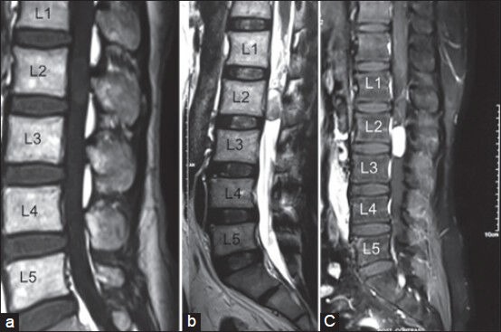
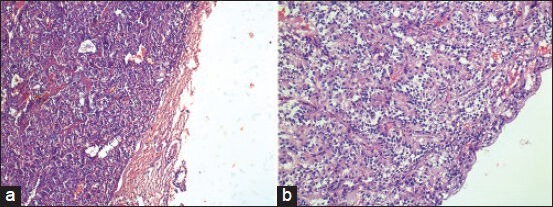
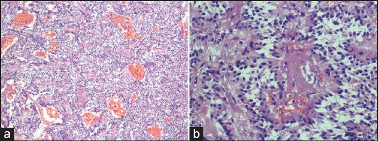
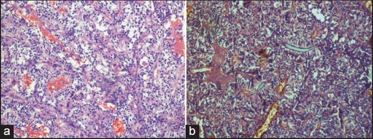
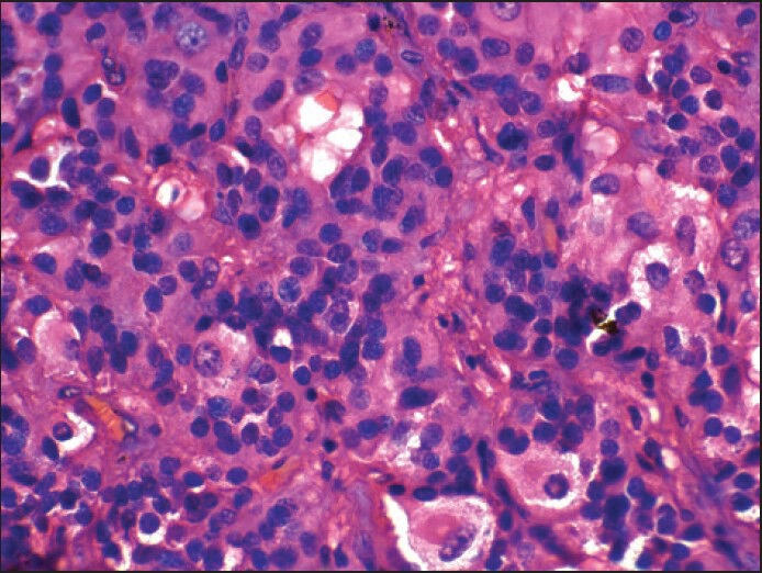
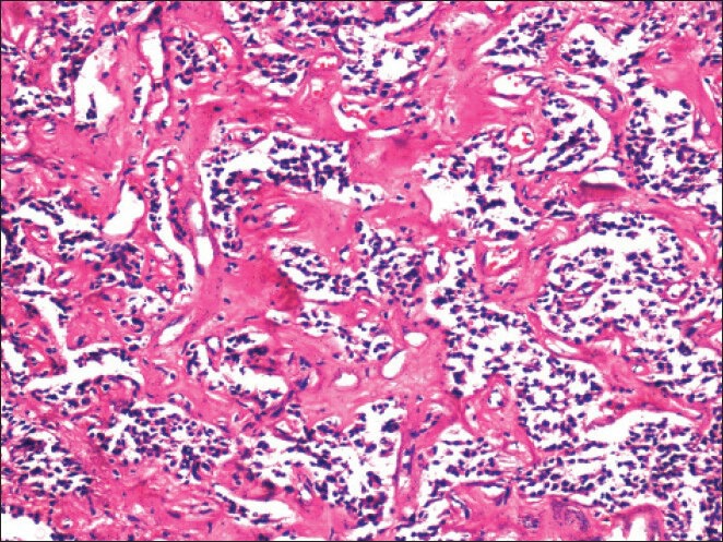
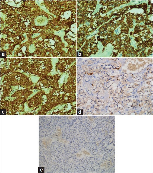
Results: There were six males and two females. The mean age at diagnosis was 50.4 years. All patients presented with low back pain. All tumors were located in the cauda equina or conus medullaris region. Magnetic Resonance Imaging and intraoperative appearance were that of a vascular, well-circumscribed intradural, extramedullary tumor suggestive of either schwannoma or ependymoma. All the patients underwent gross total resection of the tumor. Histopathology in five of the cases showed 'ependymoma-like histology' while only three cases had a predominant classic 'zellballen' pattern. Two cases had prominent 'gangliocytic differentiation'. In the five cases with 'ependymoma-like histology', the diagnosis was confirmed on Immunohistochemistry (IHC).
Conclusions: Even though relatively rare, paraganglioma should be considered in the differential diagnosis of spinal tumors and due to their clinical, radiological and histopathological similarity to schwannoma and ependymoma, the diagnosis should be based on close examination of the clinical, radiological and pathological findings.
Keywords: Cauda equina; ependymoma-like histology; paraganglioma; spinal.
Conflict of interest statement
Figures
References
-
- Lack EE. Pathology of adrenal and extra-adrenal paraganglioma. In: Livolsi VA, editor. Major Problems in Pathology. Vol. 29. Philadelphia: W.B. Saunders; 1994.
-
- Enzinger FM, Weiss SW. St. Louis: CV Mosby; 1983. Soft Tissue Tumors; pp. 676–97.
-
- Lack EE, Cubilla AL, Woodruff JM, Farr HW. Paragangliomas of the head and neck region: a clinical study of 69 patients. Cancer. 1977;39:397–409. - PubMed
-
- Kipkie GF. Simultaneous chromaffin tumors of the carotid body and the glomus jugularis. Arch Pathol (Chic) 1947;44:113–8. - PubMed
-
- Scheithauer BW, Brandner S, Soffer D. Spinal paraganglioma. In: Louis DN, Ohgaki H, Wiestler OD, Cavenee WK, editors. WHO Classification of Tumours of the Central Nervous System. Lyon: IARC; 2007. pp. 117–9.
LinkOut - more resources
Full Text Sources
Other Literature Sources