. 2014 Nov:42:169-77.
doi: 10.1016/j.bbi.2014.06.199. Epub 2014 Jul 8.

Identification of a functional interaction of HMGB1 with Receptor for Advanced Glycation End-products in a model of neuropathic pain

Affiliations

Identification of a functional interaction of HMGB1 with Receptor for Advanced Glycation End-products in a model of neuropathic pain

Yohance M Allette et al. Brain Behav Immun. 2014 Nov.

Abstract

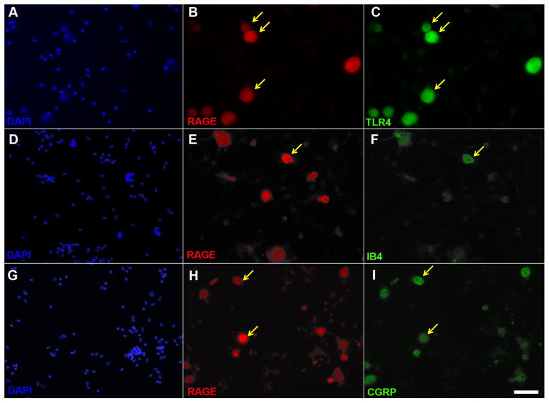
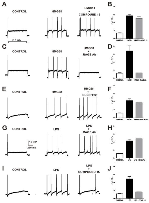
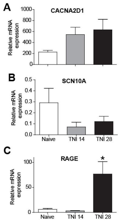
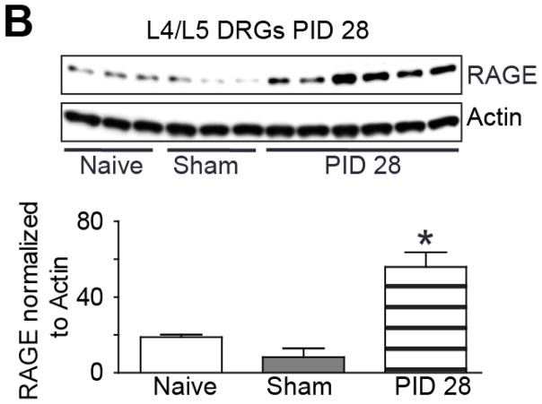
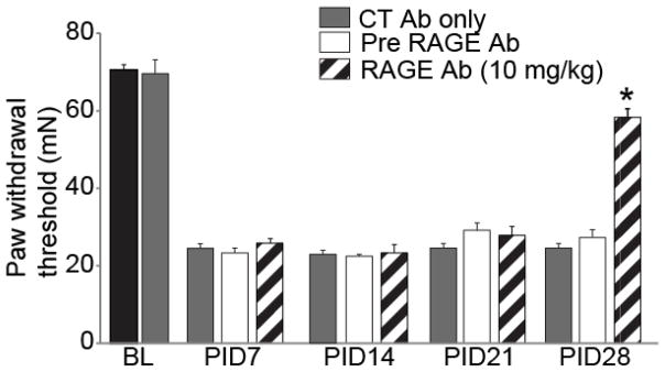
Recent studies indicate that the release of high mobility group box 1 (HMGB1) following nerve injury may play a central role in the pathogenesis of neuropathic pain. HMGB1 is known to influence cellular responses within the nervous system via two distinct receptor families; the Receptor for Advanced Glycation End-products (RAGE) and Toll-like receptors (TLRs). The degree to which HMGB1 activates a receptor is thought to be dependent upon the oxidative state of the ligand, resulting in the functional isoforms of all-thiol HMGB1 (at-HMGB1) acting through RAGE, and disufide HMGB1 (ds-HMGB1) interacting with TLR4. Though it is known that dorsal root ganglia (DRG) sensory neurons exposed to HMGB1 and TLR4 agonists can influence excitation, the degree to which at-HMGB1 signaling through neuronal RAGE contributes to neuropathic pain is unknown. Here we demonstrate that at-HMGB1 activation of nociceptive neurons is dependent on RAGE and not TLR4. To distinguish the possible role of RAGE on neuropathic pain, we characterized the changes in RAGE mRNA expression up to one month after tibial nerve injury (TNI). RAGE mRNA expression in lumbar dorsal root ganglion (DRG) is substantially increased by post-injury day (PID) 28 when compared with sham injured rodents. Protein expression at PID28 confirms this injury-induced event in the DRG. Moreover, a single exposure to monoclonal antibody to RAGE (RAGE Ab) failed to abrogate pain behavior at PID 7, 14 and 21. However, RAGE Ab administration produced reversal of mechanical hyperalgesia on PID28. Thus, at-HMGB1 activation through RAGE may be responsible for sensory neuron sensitization and mechanical hyperalgesia associated with chronic neuropathic pain states.

Keywords: Alarmin; DAMP; DRG; Dorsal root ganglia; HMGB1; Pain; RAGE; TLR4.


Conflict of interest statement

The authors declare no conflicts of interest.

Figures

Figure 1
Figure 1. RAGE, TLR4, IB4 and CGRP immunohistochemistry in primary sensory neuron cultures
Images depicting corresponding images of the cell nucleus label DAPI (A), RAGE (B) and TLR4 (C). Note that the majority of RAGE immunoreactive neurons are also positive for TLR4 (yellow arrows indicates co-expression). Corresponding images of DAPI (D), RAGE (E), and the non-peptidergic marker of nociceptive neurons, IB4 (F). A minority of IB4 cells exhibit RAGE (yellow arrow indicates co-expression). Corresponding images of DAPI (G), RAGE (H), and the peptidergic marker of nociceptive neurons, CGRP (I). Note that there are numerous RAGE immunoreactive neurons which are also positive for CGRP. Scale bar is 50 μm.
Figure 2
Figure 2. RAGE neutralizing antibody, but not the TLR4 small molecule inhibitor, Compound 15, suppresses at-HMGB1-dependent neural excitation in acutely dissociated sensory neurons
Current clamp recordings were performed on small-to-medium (>30 μm – >40 μm) diameter lumbar 4–5 DRG neurons from naive rats. Firing of 1–2 action potentials (APs) was elicited by a 1 second depolarizing current injection (ranging from 0.1 to 2.0 nA depending on the cell) every 30 seconds. Representative recordings demonstrating that application of at-HMGB1 (27μM) increases the number of elicited action potentials in DRG sensory neurons is not reversed by TLR4 small molecule inhibitor (compound 15) (A) or a TLR2 inhibitor (CU-CPT22) (E). In contrast, exposure to RAGE antibody effectively suppresses at-HMGB1-dependent action potential (C). Group data showing that compound 15 (B) and CU-CPT22 (F) do not reverse at-HMGB1-elicited increase in DRG neuron action potential firing while RAGE Ab does reverse increased excitation (D). Representative recording demonstrating that application of LPS (1 μg/ml) increases the number of elicited action potentials in DRG sensory neurons which is unaffected by RAGE antibody (G). In contrast, the neuronal effects of LPS can be suppressed with compound 15 (I). Group data showing that compound 15 (J), but not RAGE Ab (H), reverses LPS-elicited increase in DRG neuron action potential firing. (Ligand treatment compared to receptor inhibitor; *p < 0.05)
Figure 3
Figure 3. Tibal Nerve Injury (TNI) alters the expression of neuronal transcripts in dorsal root ganglion (DRG) derived from TNI rats
(A–D) RT-PCR analysis showing the mRNA expression profile of CaV alpha2delta1 (A) NaV1.8 (B) and RAGE (*p< 0.05) (C) at different time points following TNI; post injury day (PID) 14 and PID 28 (n=3). RT-PCR data were analyzed using the Ct method and mRNA expression levels are expressed relative to L27-ribosomal housekeeping gene.
Figure 4
Figure 4. RAGE protein expression following TNI at post injury day 28
Immunoblot of RAGE in L4/5 DRGs from naïve, sham injured and TNI ipsilateral to the injury at [PID] 28 (n=3, sham versus injury; *p< 0.05). Actin was used as a loading control to which samples were normalized.
Figure 5
Figure 5. Decreased tactile hyperalgesia following intraperitoneal injection of a neutralizing RAGE antibody in tibial nerve-injured rats at Day 28
Paw withdrawal threshold (PWT) in rodents subjected to TNI (n = 6–8, white bars) were significantly reduced when compared with pre-TNI thresholds (n = 6–8, black bar) for at least 28 days. Administration of control, non-neutralizing antibody did not alter PWT at baseline or after TNI at 7, 14, 21 or 28 days (n = 6–8, CT Ab; gray bars). A humanized monoclonal antibody to RAGE (RAGE Ab; 10 mg/kg body weight; n = 6–8 each) was administered intraperitoneally and ipsilateral PWT was assessed using the von Frey filament test. Behavior was tested at 1 h post injection and again at 4 hours (data not shown). A single injection of RAGE Ab did not produce a change in PWT at TNI PID 7, 14, and 21 (striped bar) that differed from pre-RAGE Ab (white bars). At TNI PID 28 a single injection of RAGE Ab successfully reversed TNI decreases in PWT when compared with pre-RAGE Ab treatment (*p< 0.05).

References

    1. Calvo M, Dawes JM, Bennett DL. The role of the immune system in the generation of neuropathic pain. Lancet Neurol. 2012;11:629–42. - PubMed
    1. Bianchi ME. DAMPs, PAMPs and alarmins: all we need to know about danger. J Leukoc Biol. 2007;81:1–5. - PubMed
    1. Andersson U, Tracey KJ. HMGB1 is a therapeutic target for sterile inflammation and infection. Annu Rev Immunol. 2011;29:139–62. - PMC - PubMed
    1. Bianchi ME, Falciola L, Ferrari S, Lilley DM. The DNA binding site of HMG1 protein is composed of two similar segments (HMG boxes), both of which have counterparts in other eukaryotic regulatory proteins. EMBO J. 1992;11:1055–63. - PMC - PubMed
    1. Czura CJ, Wang H, Tracey KJ. Dual roles for HMGB1: DNA binding and cytokine. J Endotoxin Res. 2001;7:315–21. - PubMed

Publication types

MeSH terms

Substances