. 2014 Aug 13;6(15):13209-20.
doi: 10.1021/am503043t. Epub 2014 Jul 23.

Does translational symmetry matter on the micro scale? Fibroblastic and osteoblastic interactions with the topographically distinct poly(ε-caprolactone)/hydroxyapatite thin films

Affiliations

Does translational symmetry matter on the micro scale? Fibroblastic and osteoblastic interactions with the topographically distinct poly(ε-caprolactone)/hydroxyapatite thin films

Vuk Uskoković et al. ACS Appl Mater Interfaces. .

Abstract

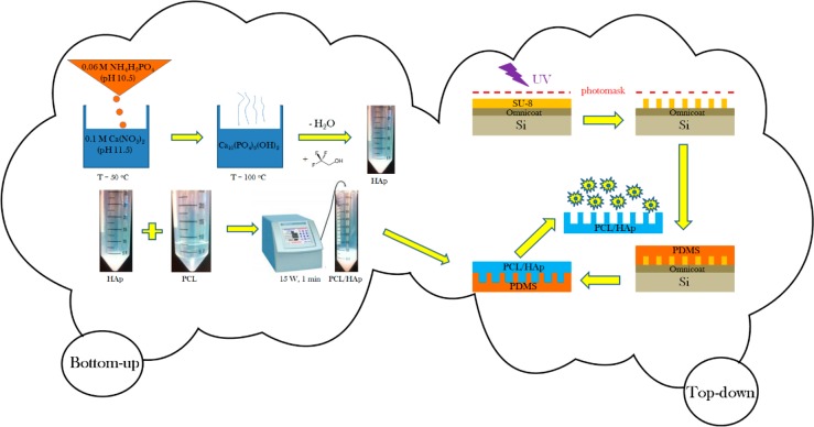
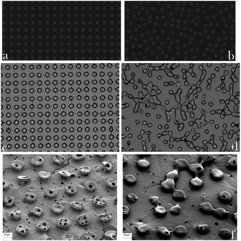
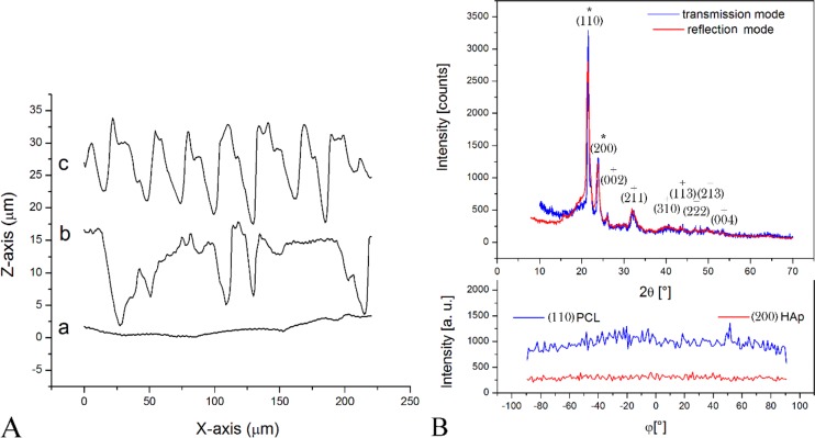
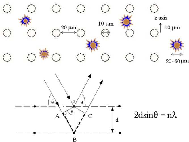
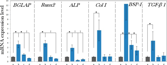
Material composition and topography of the cell-contacting material interface are important considerations in the design of biomaterials at the nano and micro scales. This study is one of the first to have assessed the osteoblastic response to micropatterned polymer-ceramic composite surfaces. In particular, the effect of topographic variations of composite poly(ε-caprolactone)/hydroxyapatite (PCL/HAp) films on viability, proliferation, migration and osteogenesis of fibroblastic and osteoblastic MC3T3-E1 cells was evaluated. To that end, three different micropatterned PCL/HAp films were compared: flat and textured, the latter of which included films comprising periodically arranged and randomly distributed oval topographic features 10 μm in diameter, 20 μm in separation and 10 μm in height, comparable to the dimensions of MC3T3-E1 cells. PCL/HAp films were fabricated by the combination of a bottom-up, soft chemical synthesis of the ceramic, nanoparticulate phase and a top-down, photolithographic technique for imprinting fine, microscale features on them. X-ray diffraction analysis indicated an isotropic orientation of both the polymeric chains and HAp crystallites in the composite samples. Biocompatibility tests indicated no significant decrease in their viability when grown on PCL/HAp films. Fibroblast proliferation and migration onto PCL/HAp films proceeded slower than on the control borosilicate glass, with the flat composite film fostering more cell migration activity than the films containing topographic features. The gene expression of seven analyzed osteogenic markers, including procollagen type I, osteocalcin, osteopontin, alkaline phosphatase, and the transcription factors Runx2 and TGFβ-1, was, however, consistently upregulated in cells grown on PCL/HAp films comprising periodically ordered topographic features, suggesting that the higher levels of symmetry of the topographic ordering impose a moderate mechanochemical stress on the adherent cells and thus promote a more favorable osteogenic response. The obtained results suggest that topography can be a more important determinant of the cell/surface interaction than the surface chemistry and/or stiffness as well as that the regularity of the distribution of topographic features can be a more important variable than the topographic features per se.


Figures

Figure 1
Figure 1
Schematic description of the fabrication of PCL/HAp films using a combination of bottom-up and top-down synthetic techniques.
Figure 2
Figure 2
AutoCAD blueprints (a, b), optical images of PDMS templates with wells on their surface (c, d) and SEM images of PCL/HAp films (e, f) with ordered (a, c, e) and disordered (b, d, f) pillar-shaped topographical features. The diameter of the spherical features is 20 μm.
Figure 3
Figure 3
(A) Profilometry diagrams of three different PCL/HAp films synthesized: (a) flat; (b) topographically disordered; (c) topographically ordered. (B) X-ray diffractograms of PCL/HAp films in transmission and reflection modes and the intensity of (110) reflection of PCL and (200) reflection of HAp in PCL/HAp films as a function of the angle, φ, denoting rotation around the axis perpendicular to the sample plane in the transmission mode. Diffraction peaks indexed with * are PCL-derived, whereas those indexed with + are HAp-derived.
Figure 4
Figure 4
Size of the surface features and the distance between them were chosen to be in the same order of magnitude as that of the size of fibroblastic cells, assuming that the surface effects on the cells would be maximized thereby, in analogy with the light diffraction process, which is most intense when the spacing between the scattering entities and the wavelength of the diffracted light are in the same range, as could be seen from Bragg’s equation, where λ is the wavelength of the incident X-ray photons, θ is the diffraction angle, d is the spacing between two nearest planes containing the scattering entities and n is the integer signifying the order constructive interference, with n = 1 being the most probable and intense one.
Figure 5
Figure 5
Temporal profile for the sustained release of fluorescein from PCL/HAp films in PBS.
Figure 6
Figure 6
(A) Proliferation of MC3T3-E1 cells after 4, 7, 10 and 12 days of incubation on different PCL/HAp films, (b) topographically ordered, (c) topographically disordered and (d) flat, normalized to proliferation on control cell culture polystyrene (a). The proliferation ratio is represented as a number of cells counted per surface area for a given sample (N) normalized to the number of cells per surface area for the control sample (Nc). (B) MTT viability assay for MC3T3-E1 cells on control cell culture polystyrene (a) and on different PCL/HAp films, (b) topographically ordered, (c) topographically disordered and (d) flat, after 1, 3, 7 and 12 days of incubation. Samples for which a statistically significant difference (p < 0.05) was observed when compared to the control are denoted with *.
Figure 7
Figure 7
Volume-rendered confocal optical micrographs of osteoblastic MC3T3-E1 cells grown for 7 days on various PCL/HAp substrates and fluorescently stained for collagen type I (red) and nucleus (blue): (a) topographically ordered, (b) topographically disordered and (c) flat.
Figure 8
Figure 8
Migration assay for MC3T3-E1 cells onto control cell culture borosilicate glass (a) and onto different PCL/HAp films, (b) topographically ordered, (c) topographically disordered and (d) flat, after 3 days of incubation. Migration density is represented as a number of cells counted per surface area for a given sample (N) normalized to the number of cells per surface area for the control sample (Nc). Statistically significant difference (p < 0.05) is denoted with *.
Figure 9
Figure 9
Normalized mRNA expression of six different osteogenic markers in MC3T3-E1 cells grown on control cell culture polystyrene (a) and on different PCL/HAp films, (b) topographically ordered, (c) topographically disordered and (d) flat, after 96 h of incubation. mRNA expression was detected by quantitative RT-polymerase chain reaction relative to the housekeeping gene β-actin (ACTB). Data normalized to the expression of ACTB are shown as averages with error bars representing standard deviation. Genes significantly (p < 0.05) upregulated with respect to the control group are marked with *. Genes significantly (p < 0.05) downregulated with respect to the control group are marked with .

Similar articles

Cited by

References

    1. Gristina A. G. Biomaterial-Centered Infection: Microbial Adhesion versus Tissue Integration. Science 1987, 237, 1588–1595. - PubMed
    1. Gu Y. X.; Du J.; Si M. S.; Mo J. J.; Qiao S. C.; Lai H. C. The Roles of PI3K/Akt Signaling Pathway in Regulating MC3T3-E1 Preosteoblast Proliferation and Differentiation on SLA and SLActive Titanium Surfaces. J. Biomed. Mater. Res., Part A 2013, 101, 748–754. - PubMed
    1. Zamani F.; Amani-Tehran M.; Latifi M.; Shokrgozar M. A. The Influence of Surface Nanoroughness of Electrospun PLGA Nanofibrous Scaffold on Nerve Cell Adhesion and Proliferation. J. Mater. Sci.: Mater. Med. 2013, 24, 1551–1560. - PubMed
    1. Teng N. C.; Wang P. D.; Chang W. J.; Feng S. W.; Fan K. H.; Lin C. T.; Hsieh S. C.; Huang H. M. Er:YAG Laser-Roughened Enamel Promotes Osteoblastic Differentiation. Photomed. Laser Surg. 2012, 30, 516–522. - PubMed
    1. Müller B. Tailoring Biocompatibility: Benefitting Patients. Mater. Today 2010, 13, 58.

Publication types

MeSH terms