. 2014 Jul 11;9(7):e102246.
doi: 10.1371/journal.pone.0102246. eCollection 2014.

Lantibiotic immunity: inhibition of nisin mediated pore formation by NisI

Affiliations

Lantibiotic immunity: inhibition of nisin mediated pore formation by NisI

Zainab AlKhatib et al. PLoS One. .

Abstract

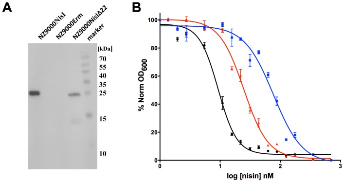
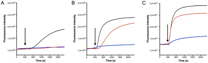
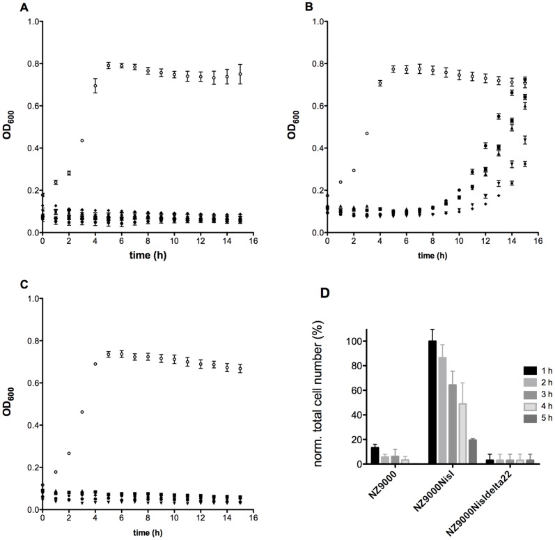
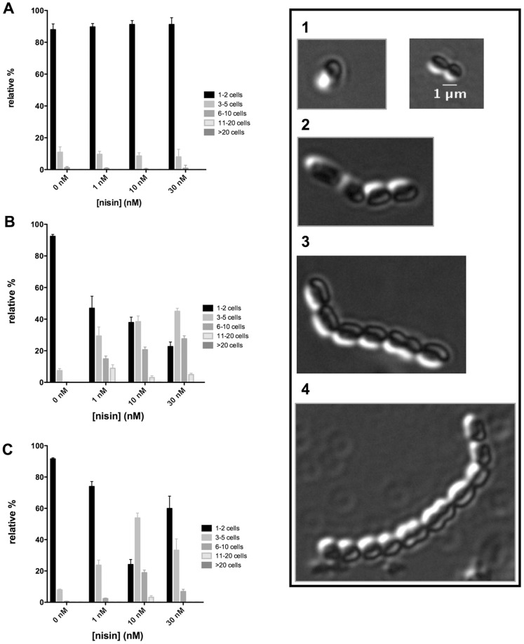
Nisin, a 3.4 kDa antimicrobial peptide produced by some Lactococcus lactis strains is the most prominent member of the lantibiotic family. Nisin can inhibit cell growth and penetrates the target Gram-positive bacterial membrane by binding to Lipid II, an essential cell wall synthesis precursor. The assembled nisin-Lipid II complex forms pores in the target membrane. To gain immunity against its own-produced nisin, Lactococcus lactis is expressing two immunity protein systems, NisI and NisFEG. Here, we show that the NisI expressing strain displays an IC50 of 73 ± 10 nM, an 8-10-fold increase when compared to the non-expressing sensitive strain. When the nisin concentration is raised above 70 nM, the cells expressing full-length NisI stop growing rather than being killed. NisI is inhibiting nisin mediated pore formation, even at nisin concentrations up to 1 µM. This effect is induced by the C-terminus of NisI that protects Lipid II. Its deletion showed pore formation again. The expression of NisI in combination with externally added nisin mediates an elongation of the chain length of the Lactococcus lactis cocci. While the sensitive strain cell-chains consist mainly of two cells, the NisI expressing cells display a length of up to 20 cells. Both results shed light on the immunity of lantibiotic producer strains, and their survival in high levels of their own lantibiotic in the habitat.


Conflict of interest statement

Competing Interests: The authors have declared that no competing interests exist.

Figures

Figure 1
Figure 1. Western blot analysis using a polyclonal NisI antibody (A).
Shown are the L. lactis strains: NZ9000Erm, NZ9000NisI and NZ9000NisIΔ22 strain. IC50 determination of different strains (B). Growth inhibition experiments were performed with nisin using different strains. Black line: NZ9000Erm strain; blue line: NZ9000NisI strain; red line: NZ9000NisIΔ22 strain. Data was fitted and evaluated according to equation (1). Each experiment was performed at least in triplicates.
Figure 2
Figure 2. SYTOx green assay to visualize pore formation mediated by nisin.
The NZ9000Erm strain (black line), NZ9000NisI strain (blue line) and the NZ9000NisIΔ22 strain (red line) were grown and incubated with the SYTOX green dye. The fluorescence signal was monitored online using a fluorolog (Horiba III). After 400 seconds a stable baseline was reached and nisin was added (A) 10 nM (B) 30 nM and (C) 1000 nM. The addition of nisin is indicated with an arrow. The rapid increase of the fluorescence signal indicated pore formation. The data are representatives of at least three independent measurements.
Figure 3
Figure 3. Growth recovery assay.
The different strains were incubated for 1 (•), 2 (▪), 3(▴), 4(▾) and 5(⧫) hours at an OD600 of 0.1 with nisin at a concentration which represents 10-fold the IC50 determined, 100 nM, 300 nM and 600 nM for the NZ9000Erm (A), NZ9000NisIΔ22 (B) and the NZ9000NisI (C) strains, respectively. The cells were separated from the growth media by centrifugation and extensively washed with media to remove the remaining nisin. Afterwards the cells were transferred into fresh medium at a final OD600 of 0.1 and the growth was monitored by measuring the optical density at 600 nm. As a control (○) the corresponding strains without the addition of nisin during pre-incubation were used. Each experiment was performed 4 times. Within the different experiments, the interval of recovery comprised between 5 and 8 hours. Furthermore the end point OD600 (after 15 hours growth) was in a range of 65–100% recovery ability (compared with the end point OD600 of the control). To control the number of cells surviving the incubation with high nisin concentrations, the resuspended cells were striked out on GM17 agar plates. The number of colonies on these plates resemble the total number of living cell in the cell suspension with an OD600 of 0.1. A normalisation of the total cell number between the strains NZ9000Erm, NZ9000NisI and NZ9000NisIΔ22 shows the relative distribution depending on the living cells (D). The NZ9000 NisI after 1 h incubation time is set as 100% (total cell number: 261.866±32.809) and reflects the 1.4% of surviving cells compared to the control (total cell number: 24.800.000±1.844.776). Longer incubation times lead to survival rates of 20% for NZ9000NisI. Even fewer cells, only 0.1%, survived for the NZ9000Erm and NZ9000NisIΔ22 strains, when compared to the control. The error bars indicating the standard deviation of three independent experiments.
Figure 4
Figure 4. Phenotype visualisation of L. lactis cells using the NZ9000Erm, NZ9000NisI and NZ9000NisIΔ22 strain.
The different strains were grown until exponential phase (OD600 = 0.5). During growth, different concentrations of nisin were added (0, 1, 10 and 30 nM). The cells were transferred and fixed onto a coverslide and the cells were visualised using a Nikon eclipse Ti microscope. The chains were counted and categorized in different classes. Class 1 consisted of 1–2 cocci (black bar), class 2 consisted of 3–5 cells (grey bar), class 3 consisted of 6–10 cells (dark grey bar), class 4 comprised of 11–20 cells (light grey) and class 5 comprised of >20 cells. For each sample the number of counted cells per area was >50. In total, after scanning five different areas at least >500 cell chains were observed.

References

    1. Cleveland J, Montville TJ, Nes IF, Chikindas ML (2001) Bacteriocins: safe, natural antimicrobials for food preservation. Int J Food Microbiol 71: 1–20. - PubMed
    1. Tagg JR, Dajani AS, Wannamaker LW (1976) Bacteriocins of gram-positive bacteria. Bacteriol Rev 40: 722–756. - PMC - PubMed
    1. Cotter PD, Hill C, Ross RP (2005) Bacteriocins: developing innate immunity for food. Nat Rev Microbiol 3: 777–788. - PubMed
    1. Kruszewska D, Sahl HG, Bierbaum G, Pag U, Hynes SO, et al. (2004) Mersacidin eradicates methicillin-resistant Staphylococcus aureus (MRSA) in a mouse rhinitis model. J Antimicrob Chemother 54: 648–653. - PubMed
    1. Galvin M, Hill C, Ross RP (1999) Lacticin 3147 displays activity in buffer against gram-positive bacterial pathogens which appear insensitive in standard plate assays. Lett Appl Microbiol 28: 355–358. - PubMed

Publication types

MeSH terms

Substances