. 2014 Jul 11;9(7):e100230.
doi: 10.1371/journal.pone.0100230. eCollection 2014.

Ceftriaxone preserves glutamate transporters and prevents intermittent hypoxia-induced vulnerability to brain excitotoxic injury

Affiliations

Ceftriaxone preserves glutamate transporters and prevents intermittent hypoxia-induced vulnerability to brain excitotoxic injury

Rekha Jagadapillai et al. PLoS One. .

Abstract

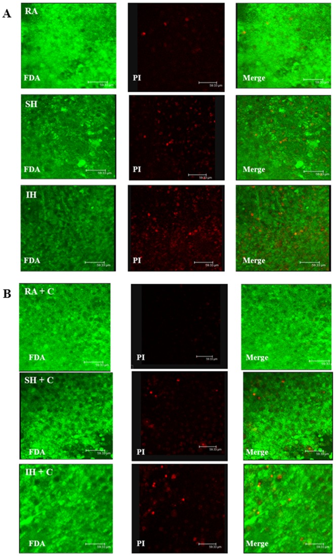
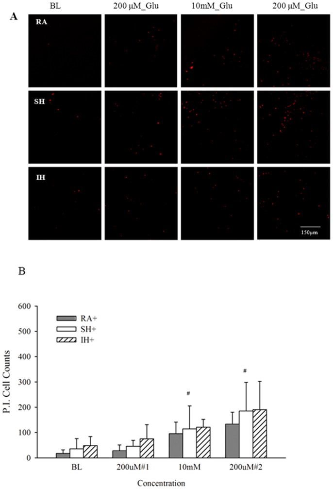
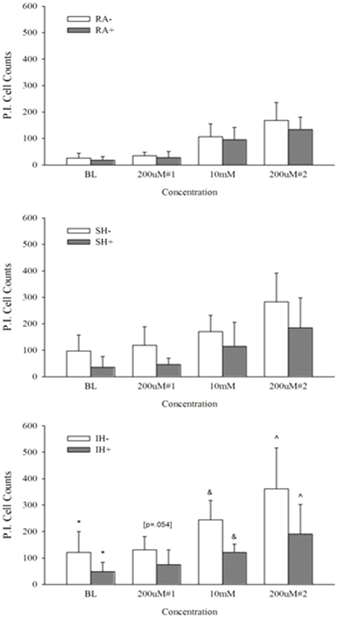
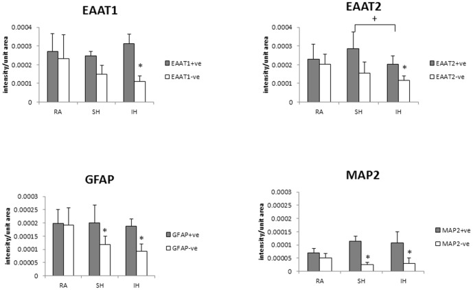
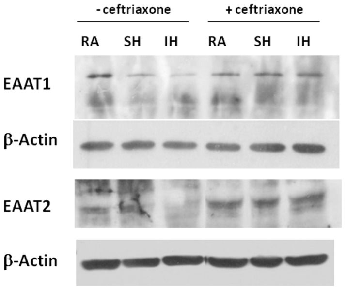
Hypoxia alters cellular metabolism and although the effects of sustained hypoxia (SH) have been extensively studied, less is known about chronic intermittent hypoxia (IH), commonly associated with cardiovascular morbidity and stroke. We hypothesize that impaired glutamate homeostasis after chronic IH may underlie vulnerability to stroke-induced excitotoxicity. P16 organotypic hippocampal slices, cultured for 7 days were exposed for 7 days to IH (alternating 2 min 5% O2-15 min 21% O2), SH (5% O2) or RA (21% O2), then 3 glutamate challenges. The first and last exposures were intended as a metabolic stimulus (200 µM glutamate, 15 min); the second emulated excitotoxicity (10 mM glutamate, 10 min). GFAP, MAP2, and EAAT1, EAAT2 glutamate transporters expression were assessed after exposure to each hypoxic protocol. Additionally, cell viability was determined at baseline and after each glutamate challenge, in presence or absence of ceftriaxone that increases glutamate transporter expression. GFAP and MAP2 decreased after 7 days IH and SH. Long-term IH but not SH decreased EAAT1 and EAAT2. Excitotoxic glutamate challenge decreased cell viability and the following 200 µM exposure further increased cell death, particularly in IH-exposed slices. Ceftriaxone prevented glutamate transporter decrease and improved cell viability after IH and excitotoxicity. We conclude that IH is more detrimental to cell survival and glutamate homeostasis than SH. These findings suggest that impaired regulation of extracellular glutamate levels is implicated in the increased brain susceptibility to excitotoxic insult after long-term IH.


Conflict of interest statement

Competing Interests: The authors have declared that no competing interests exist.

Figures

Figure 1
Figure 1. Long term sustained or Intermittent hypoxia decreases cell viability.
Propidium iodide (red) and Fluorescein diacetate (green) staining of slices exposed to 7 days RA, SH or IH (A) without and (B) with 100 µM ceftriaxone (n = 4-6).
Figure 2
Figure 2. Long term sustained or Intermittent hypoxia decreases cell viability.
LDH release in the supernatants of slices exposed to 7 days RA, SH or IH with (+) or without 100 µM ceftriaxone, presented as mean + SEM (n = 5–9). * RA<SH (p = 0.01). * RA<IH (p = 0.02).
Figure 3
Figure 3. Intermittent hypoxia decreases cell viability and impairs glutamate response.
Propidium iodide staining (A) and quantification of PI positive cells presented as mean + SD (B) of slices exposed to 7 days RA, SH or IH at baseline (BL), after 200 µM glutamate, and 10 mM glutamate, followed by a second 200 µM glutamate challenge. n = 12–18. *: At all concentrations RA- < SH- & IH- (p<.001). + At 10mM: SH- < IH- (p<.05). # At 10 mM & 200 µM#2: BL < SH- & IH- (p<.05 and p<.001 respectively).
Figure 4
Figure 4. Ceftriaxone prevents hypoxia-induced cell death and improves tolerance to excitotoxicity.
Propidium iodide staining (A) and quantification of PI positive cells presented as mean + SD (B) of slices exposed to 100 µM ceftriaxone during 7 days RA, SH or IH at baseline (BL), after 200 µM glutamate, and 10 mM glutamate, followed by a second 200 µM glutamate challenge. baseline (BL) n = 9–12, # At 10 mM & 200 µM#2: RA+; IH+; SH+> their respective BL (p<.01).
Figure 5
Figure 5. Ceftriaxone effect on cell tolerance to glutamate is significantly greater in IH-exposed slices.
Propidium iodide staining quantification of ceftriaxone (+) treated slices exposed to RA, SH and IH at baseline (BL), after 200 µM glutamate, and 10 mM glutamate, followed by a second 200 µM glutamate challenge. n = 12–18 for RA-, SH-, IH- and n = 9-12 for RA+, SH+, IH+; IH- > IH+: *At BL (p = .005), &At 10 mM (p<.001) & ∧At 200 µM#2 (p = .005).
Figure 6
Figure 6. IH significantly decreases glutamate transporters, MAP2 and GFAP immunoreactivity.
EAAT1, EAAT2, GFAP and MAP2 immunoreactivity in slices exposed to 7 days RA, SH or IH. EAAT1 and EAAT2 expression was unchanged by SH while significantly reduced in IH. MAP2 and GFAP expression decreased in both SH and IH.
Figure 7
Figure 7. Ceftriaxone prevents IH-induced decrease in glutamate transporters, MAP2 and GFAP immunoreactivity.
EAAT1, EAAT2, GFAP and MAP2 immunoreactivity in slices exposed to 7 days RA, SH or IH in presence of 100 µM ceftriaxone. Ceftriaxone treatment prevented the decrease in EAAT1, EAAT2 in IH, and of MAP2 and GFAP in both IH and SH.
Figure 8
Figure 8. Graphic representation of glutamate transporters, MAP2 and GFAP immunoreactivity with or without ceftriaxone.
Quantification of EAAT1, EAAT2, GFAP and MAP2 immunofluorescence per unit area of slices exposed to 7 days RA, SH or IH in presence (+ve) or in absence (-ve) of 100 µM ceftriaxone. Data are presented as mean immunofluorescence + SD. n =  4-13 for RA+, IH+, SH+; n = 5–11 for RA-, IH-, SH *: IH- or SH- <RA- (p≤0.01). +: IH+ < SH+ (p≤0.01).
Figure 9
Figure 9. EAAT1 and EAAT2 immunoblotting of slices exposed to RA, SH and IH with or without ceftriaxone shows that ceftriaxone prevents IH-induced decrease in glutamate transporters.
EAAT1 and EAAT2, immunoblotting of lysates from slices exposed to 7 days RA, SH or IH in absence or in presence of 100 µM ceftriaxone. Glutamate transporters expression decreased in IH-exposed slices. Ceftriaxone treatment increased EAAT1 and EAAT2 expression in all conditions and prevented EAAT1 and EAAT2 decrease in IH.

References

    1. Schurr A, Miller JJ, Payne RS, Rigor BM (1999) An increase in lactate output by brain tissue serves to meet the energy needs of glutamate-activated neurons. J Neurosci 19: 34–39. - PMC - PubMed
    1. Vega C, Sachleben R, Gozal D, Gozal E (2006) Differential metabolic adaptation to acute and long-term hypoxia in rat primary cortical astrocytes. J Neurochem 97: 872–883. - PubMed
    1. Yaggi HK, Concato J, Kernan WN, Lichtman JH, Brass LM, et al. (2005) Obstructive sleep apnea as a risk factor for stroke and death. N Engl J Med 353: 2034–2041. - PubMed
    1. Arzt M, Young T, Finn L, Skatrud JB, Bradley TD (2005) Association of sleep-disordered breathing and the occurrence of stroke. Am J Respir Crit Care Med 172: 1447–1451. - PMC - PubMed
    1. Yan-fang S, Yu-ping W (2009) Sleep-disordered breathing: impact on functional outcome of ischemic stroke patients. Sleep Med 10: 717–719. - PubMed

Publication types

MeSH terms

LinkOut - more resources