Review
. 2014 Jul 11:16:103-29.
doi: 10.1146/annurev-bioeng-071813-104733.

Optical neural interfaces

Affiliations
Review

Optical neural interfaces

Melissa R Warden et al. Annu Rev Biomed Eng. .

Abstract

Genetically encoded optical actuators and indicators have changed the landscape of neuroscience, enabling targetable control and readout of specific components of intact neural circuits in behaving animals. Here, we review the development of optical neural interfaces, focusing on hardware designed for optical control of neural activity, integrated optical control and electrical readout, and optical readout of population and single-cell neural activity in freely moving mammals.

Keywords: GCaMP; channelrhodopsin; halorhodopsin; imaging; neurophysiology; optogenetics.


Figures

Figure 1
Figure 1
Optogenetics for control of genetically or topologically defined neural subtypes. (a) Electrical stimulation can be effective for modulating local neural activity, but the heterogeneity of brain tissue precludes control of single cell types using this method (left). Genetically targetable optogenetic constructs enable more precise stimulation of specified neural subtypes intermingled with nontargeted neurons (right). (Panel modified with permission from 154.) (b) Many different types of optogenetic constructs have been developed for control of neural activity. Currently available tools allow for induction of action potentials (ChR-family light-gated cation channels shown, left), silencing of neural activity (HR-family light-gated chloride pumps shown, middle; proton pumps may also be used), and modulation of intracellular signaling cascades [OptoXR family (153) shown, right; other classes of biochemical tool may also be used], among many other applications. (Panel modified with permission from 4.) Abbreviations: ChR, channelrhodopsin; ChR2, channelrhodopsin-2; HR, halorhodopsin; NpHR, halorhodopsin from Natronomonas pharaonis; optoXR, opsin-receptor chimaera.
Figure 2
Figure 2
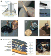
Optical-neural interfaces for light delivery to brain tissue in freely moving mammals. (a) The original fiberoptic neural interface (27a), in this case implemented via a cannula implanted over the brain region of interest; the optical fiber coupled to a light source is inserted during behavioral testing. This approach allows for simple integration with pharmacological methods. (Panel modified with permission from .) (b) Light delivery through an implanted fiber. An optical fiber stub is implanted over the brain region of interest and is coupled to a fiberoptic tether with a ceramic sheath during behavior. This approach is well suited to high-throughput behavioral testing but cannot be easily combined with pharmacology at the same site. Both cannulas and optical fiber stubs can be used for single- or dual-site illumination. (Panel modified with permission from 29 and 154.) Further advancements include (c) multisite light delivery (panel modified with permission from 42) and (d) wireless control (panel modified with permission from 37), both appropriately sized for use in freely behaving mice. (e) Injectable optoelectronics for wireless multisite multiwavelength optical stimulation/sensing, electrophysiology, and temperature sensing with coregistration and minimal tissue damage. Multiple μ-ILED light sources (a thousandth the size of conventional LEDs) can be independently controlled and can deliver a choice of wavelengths. Abbreviations: μ-IPD, microscale inorganic photodetector; μ-ILED, microscale inorganic light-emitting diode. (Panel modified with permission from 48.)
Figure 3
Figure 3
Integrated neurophysiology and optical control. (a) Optical fibers can be incorporated into multisite silicon probe designs suitable for use in freely behaving rats. (Panel modified with permission from 58.) (b) A tetrode microdrive with independently drivable tetrodes can be modified to incorporate either a fixed or freely movable optical fiber for use in large, superficial brain regions such as cortex or the hippocampus. (Panel modified with permission from 63.) (c) Simultaneous optical stimulation and electrical recording from small, deep structures, such as the brainstem neuromodulatory nuclei, requires a more compact design. A drivable device achieves this with tetrodes attached to a central fiber core. (Panel modified with permission from 61.)(d) An optical fiber can replace an electrode on a multielectrode array to enable simultaneous optical stimulation and high-density cortical recording. (Panel modified with permission from 65.)
Figure 4
Figure 4
Optical-neural interfaces for readout of neural signals. Several approaches have been developed for optically monitoring neural signals in freely behaving mammals. (a) Fiber-based bulk tissue optical recording. A single multimode optical fiber is implanted over a brain area of interest in order to record fluorescence signals from neurons expressing genetically encoded Ca2+ or voltage indicators. This system is particularly suitable for use in freely behaving mice owing to the small form factor and minimal weight of the implanted fiber. (Panel modified with permission from 96.) (b) One-photon imaging through a fiber bundle permits the construction of population-scale two-dimensional images, which can be used to monitor layer-specific neural activity. (Panel modified with permission from .)(c) Head-mounted two-photon microscopy suitable for imaging cellular-resolution cortical fluorescence signals in behaving rats (97). (d) A lightweight one-photon endoscopic microscopy system that can be used to image cellular-resolution neural activity in deep structures in behaving mice. (Panel modified with permission from 101.) (e) Ca2+-imaging data collected with the system depicted in panel d. (top) CA1 principal neurons identified by Ca2+ imaging. (bottom) Ca2+ signals recorded from this neural population in a freely behaving mouse. (Panel modified with permission from 146.) Abbreviations: CMOS, complementary metal-oxide semiconductor; F, fluorescence; LED, light-emitting diode; OGB-1 AM, Oregon Green 488 BAPTA-1; PMT, photomultiplier tube; ROI, region of interest; TCSPC, time-correlated single-photon counting.

References

    1. Penfield W, Perot P. The brain’s record of auditory and visual experience: a final summary and discussion. Brain. 1963;86(4):595–696. - PubMed
    1. Salzman CD, Britten KH, Newsome WT. Cortical microstimulation influences perceptual judgements of motion direction. Nature. 1990;346(6280):174–77. - PubMed
    1. Mayberg HS, Lozano AM, Voon V, McNeely HE, Seminowicz D, et al. Deep brain stimulation for treatment-resistant depression. Neuron. 2005;45(5):651–60. - PubMed
    1. Fenno L, Yizhar O, Deisseroth K. The development and application of optogenetics. Annu Rev Neurosci. 2011;34:389–412. - PMC - PubMed
    1. Yizhar O, Fenno LE, Davidson TJ, Mogri M, Deisseroth K. Optogenetics in neural systems. Neuron. 2011;71(1):9–34. - PubMed

Publication types