

Physical properties and biological/odontogenic effects of an experimentally developed fast-setting α-tricalcium phosphate-based pulp capping material
- PMID: 25015173
- PMCID: PMC4105101
- DOI: 10.1186/1472-6831-14-87
Physical properties and biological/odontogenic effects of an experimentally developed fast-setting α-tricalcium phosphate-based pulp capping material
Abstract
Background: Recently, fast-setting α-tricalcium-phosphate (TCP) cement was developed for use in the pulp capping process. The aim of this study was to investigate the physical properties and biological effects of α-TCP cement in comparison with mineral trioxide aggregate (MTA).
Methods: We measured the setting time, pH values, compressive strength, and solubility of the two materials. We evaluated biocompatibility on the basis of cell morphology and a viability test using human dental pulp cells (hDPCs). Chemical composition of each material was analyzed by energy dispersive x-ray spectroscopic (EDS) analysis. The expression of odontogenic-related genes was evaluated by Western blotting and immunofluorescence. The calcified nodule formation was measured by Alizarin red staining. We performed the pulp capping procedure on rat teeth for histological investigation. The data were analyzed by an independent t-test for physical properties, one-way ANOVA for biological effects, and the Mann-Whitney U test for tertiary dentin formation. A P value of less than 0.05 was considered statistically significant for all tests.
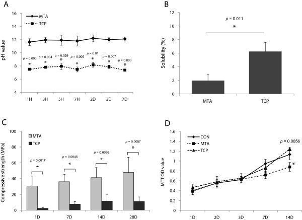
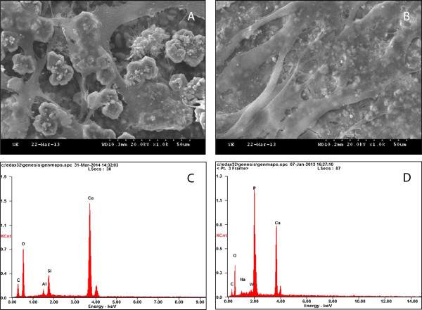
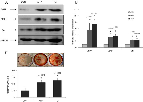
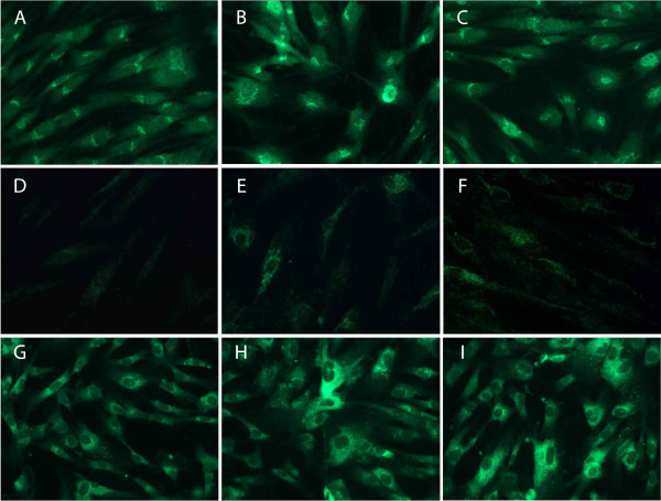
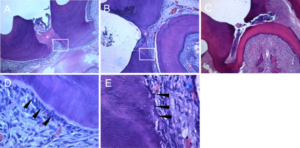
Results: The setting time, pH values, and compressive strength of α-TCP was lower than that of MTA (P < 0.05); however, the solubility of α-TCP was higher than that of MTA (P < 0.05). The resultant cell viability observed with the two materials was similar (P > 0.05). Scanning electron microscopy (SEM) revealed that cells attached to both materials were flat and had cytoplasmic extensions. The expression of odontogenic-related markers and mineralized nodule formation were higher in the two experimental groups compared to the control group (P < 0.05). Continuous tertiary dentin was formed underneath the capping materials in all samples of the tested groups.
Conclusions: Our study demonstrated that the α-TCP exhibited biocompatibility and odontogenicity comparable to MTA, whereas it had a quicker setting time.
Figures
References
-
- Kurashina K, Kurita H, Hirano M, Kotani A, Klein CP, de Groot K. In vivo study of calcium phosphate cements: Implantation of an alpha-tricalcium phsphate/dicalcium phosphate dibasic/tetracalcium phosphate monoxide cement paste. Biomaterials. 1997;18:539–549. - PubMed
-
- Blom EJ, Klein-Nulend J, Wolke JG, Kurashina K, van Waas MA, Burger EH. Transforming growth factor-beta1 incorporation in an alpha-tricalcium phosphate/dicalcium phosphate dihydrate/tetracalcium phosphate monoxide cement: release characteristics and physicochemical properties. Biomaterials. 2002;23:1261–1268. - PubMed
-
- Yoshimine Y, Akamine A, Mukai M, Maeda K, Matsukura M, Kimura Y, Makishima T. Biocompatibility of tetracalcium phosphate cement when used as a bone substitute. Biomaterials. 1993;14:403–406. - PubMed
-
- Rajesh JB, Nandakumar K, Varma HK, Komath M. Calcium phosphate cement as a "barrier-graft" for the treatment of human periodontal intraosseous defects. Indian J Dent Res. 2009;20:471–479. - PubMed
-
- Baldock WT, Hutchens LH Jr, McFall WT Jr, Simpson DM. An evaluation of tricalcium phosphate implants in human periodontal osseous defects of two patients. J Periodontol. 1985;56:1–7. - PubMed
Publication types
MeSH terms
Substances
LinkOut - more resources
Full Text Sources
Other Literature Sources
