Review
. 2015 Apr;30(4):575-83.
doi: 10.1093/ndt/gfu230. Epub 2014 Jul 12.

Cell cycle arrest and the evolution of chronic kidney disease from acute kidney injury

Affiliations
Review

Cell cycle arrest and the evolution of chronic kidney disease from acute kidney injury

Guillaume Canaud et al. Nephrol Dial Transplant. 2015 Apr.

Abstract

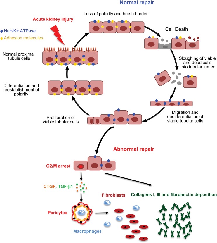
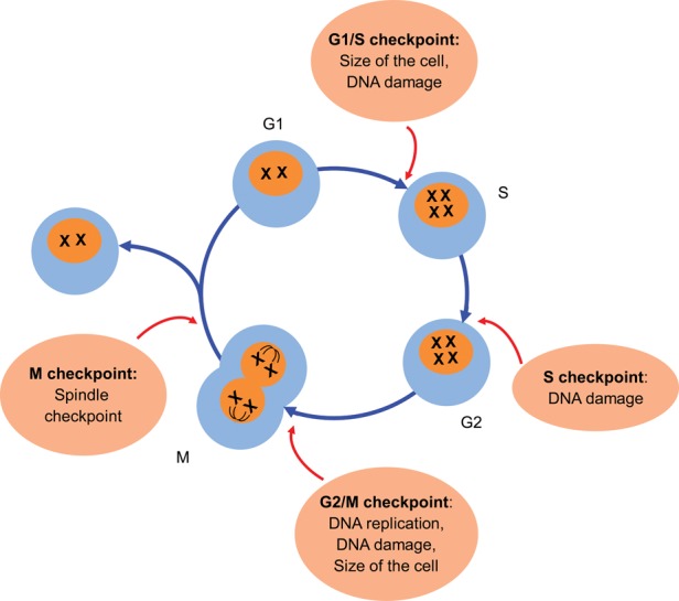
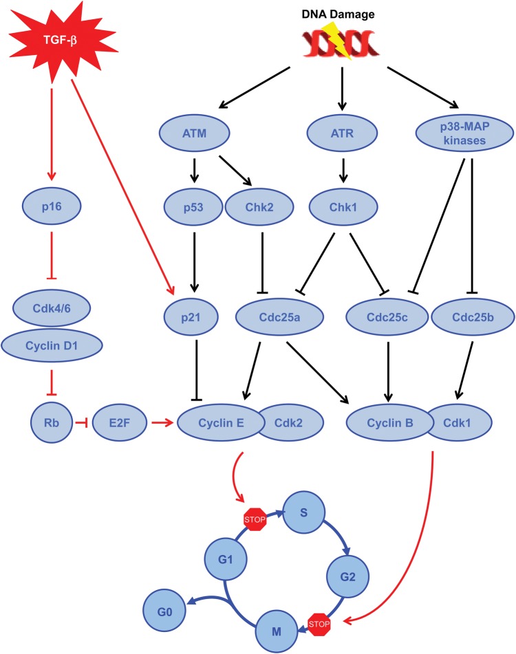
For several decades, acute kidney injury (AKI) was generally considered a reversible process leading to complete kidney recovery if the individual survived the acute illness. Recent evidence from epidemiologic studies and animal models, however, have highlighted that AKI can lead to the development of fibrosis and facilitate the progression of chronic renal failure. When kidney injury is mild and baseline function is normal, the repair process can be adaptive with few long-term consequences. When the injury is more severe, repeated, or to a kidney with underlying disease, the repair can be maladaptive and epithelial cell cycle arrest may play an important role in the development of fibrosis. Indeed, during the maladaptive repair after a renal insult, many tubular cells that are undergoing cell division spend a prolonged period in the G2/M phase of the cell cycle. These tubular cells recruit intracellular pathways leading to the synthesis and the secretion of profibrotic factors, which then act in a paracrine fashion on interstitial pericytes/fibroblasts to accelerate proliferation of these cells and production of interstitial matrix. Thus, the tubule cells assume a senescent secretory phenotype. Characteristic features of these cells may represent new biomarkers of fibrosis progression and the G2/M-arrested cells may represent a new therapeutic target to prevent, delay or arrest progression of chronic kidney disease. Here, we summarize recent advances in our understanding of the biology of the cell cycle and how cell cycle arrest links AKI to chronic kidney disease.


Figures

FIGURE 1:
FIGURE 1:
Normal and abnormal repair after AKI. After injury, tubular cells, and especially proximal tubular cells, lose their polarity and brush border; membrane proteins and tubule cells die if the injury is sustained. During the normal process of repair after AKI, surviving tubular cells undergo dedifferentiation, then migrate along the basement membrane, proliferate and finally differentiate to restore a functional nephron. However, in some conditions, the recovery process after injury becomes maladaptive and AKI leads to abnormal repair with persistent parenchyma inflammation, fibroblast proliferation and excessive deposition of extracellular matrix. CTGF, connective tissue growth factor; TGF-β1, transforming growth factor beta-1.
FIGURE 2:
FIGURE 2:
Cell cycle and checkpoints. Cell division is the result of a tightly regulated sequence of events leading to the birth of two daughter cells. It consists of four distinct phases with specific changes (G0–G1, S, G2 and M). The progression through the cell cycle is controlled by cyclic proteins, called cyclins, cyclin-dependent kinases and their inhibitors.
FIGURE 3:
FIGURE 3:
Pathways involved to block cell cycle progression. Brief summary of the different pathways involved to block cell cycle progression during the G2/M phase, including either DNA damage (black arrows) or cytokine pathway (red arrows). TGF-β, transforming growth factor beta; ATM, ataxia telangiectasia mutated; ATR, ataxia telangiectasia and Rad3-related; Chk, checkpoint kinase; Cdc, cell division cycle; MAP kinases, mitogen-activated protein kinases; CDK, cyclin-dependent kinases; Rb, retinoblastoma protein.
FIGURE 4:
FIGURE 4:
Cell cycle distribution of tubular cells in different models of AKI. Cell cycle distribution (G1, S and G2/M) of tubular cells in moderate ischemia–reperfusion injury (IRI) (a), unilateral ischemia reperfusion injury (UIRI) (b) and acute aristolochic acid toxic nephropathy (AAN) (c) models as a function of time after the insult. Extract from [2].

References

    1. Basile DP, Donohoe D, Roethe K, et al. Renal ischemic injury results in permanent damage to peritubular capillaries and influences long-term function. Am J Physiol Renal Physiol 2001; 281: F887–F899 - PubMed
    1. Yang L, Besschetnova TY, Brooks CR, et al. Epithelial cell cycle arrest in G2/M mediates kidney fibrosis after injury. Nat Med 2010; 16: 535–543 - PMC - PubMed
    1. Hsu CY. Yes, AKI truly leads to CKD. J Am Soc Nephrol 2012; 23: 967–969 - PubMed
    1. Coca SG, Singanamala S, Parikh CR. Chronic kidney disease after acute kidney injury: a systematic review and meta-analysis. Kidney Int 2012; 81: 442–448 - PMC - PubMed
    1. Khosla N, Soroko SB, Chertow GM, et al. Preexisting chronic kidney disease: a potential for improved outcomes from acute kidney injury. Clin J Am Soc Nephrol 2009; 4: 1914–1919 - PMC - PubMed

Publication types

MeSH terms