Review
. 2014 Dec 15:79-80:68-78.
doi: 10.1016/j.addr.2014.07.002. Epub 2014 Jul 10.

Microfluidic 3D models of cancer

Affiliations
Review

Microfluidic 3D models of cancer

Kyung Eun Sung et al. Adv Drug Deliv Rev. .

Abstract

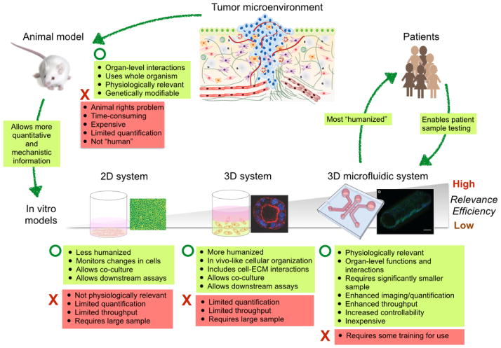
Despite advances in medicine and biomedical sciences, cancer still remains a major health issue. Complex interactions between tumors and their microenvironment contribute to tumor initiation and progression and also contribute to the development of drug resistant tumor cell populations. The complexity and heterogeneity of tumors and their microenvironment make it challenging to both study and treat cancer. Traditional animal cancer models and in vitro cancer models are limited in their ability to recapitulate human structures and functions, thus hindering the identification of appropriate drug targets and therapeutic strategies. The development and application of microfluidic 3D cancer models have the potential to overcome some of the limitations inherent to traditional models. This review summarizes the progress in microfluidic 3D cancer models, their benefits, and their broad application to basic cancer biology, drug screening, and drug discovery.

Keywords: 3D in vitro system; Biomimetics; Cancer; Drug testing; High-throughput screening; Microfluidics; Tumor microenvironment.


Figures

Fig. 1
Fig. 1
Comparison of existing cancer research models. The illustration of tumor microenvironment is adopted from Ref. [100] and the microfluidic channel image is reproduced from Ref. [59] with permission.
Fig. 2
Fig. 2
Representative microfluidic 3D systems. (a) schematic illustration of tumor spheroids generated in microfabricated well. Reproduced from Ref. [34] with permission. (b) Illustration of he hanging drop array plate to culture 3D spheroids. The 384 hanging drop array plate is sandwiched between a 96-well plate filled with distilled water and a standard-sized plate lid. Distilled water from the bottom 96-well plate and the peripheral water reservoir prevent serious evaporation of the small volume hanging drops. Reproduced from Ref. [36] with permission. (c) 3D micro-compartmentalization driven by laminar flow in microchannels. Reproduced from Ref. [40] with permission. Drops of cell containing polymer solutions are loaded onto the inlet ports. Laminar flow leads to two side-by-side 3D compartments. (d) Schematic diagram of the microfluidic gradient device used for photopolymerization of hydrogels. Reproduced from Ref. [101] with permission. the device consists of a pat- terned PDMS mold attached to an activated glass slide. All of the inlets are filled with a solution containing 8 % acrylamide and the photoinitiator. To generate a gradient in the crosslinker concentration, 0.04 % bis was added to two adjacent inlets, and 0.48 % bis to the third inlet.
Fig. 3
Fig. 3
Three-dimensional formation of endothelial sprouts and neovessels in a microfluidic device. (A) Device schematic. Parallel cylindrical channels are encased in a 3D collagen matrix within a microfabricated PDMS gasket and connected to fluid reservoirs. One channel is coated with endothelial cells and perfused with medium and the other channel is perfused with medium enriched with angiogenic factors. (B) Photograph of the device. Zoom shows phase (Upper) and fluorescent (Lower) micrographs of an endothelialized channel. F-actin and nuclei are labeled with phalloidin (green) and DAPI (blue), respectively. (C) Representative confocal immunofluorescence images of sprouting and migrating endothelial cells in response to gradients of different proangiogenic factors. Reproduced from Ref. [102] with permission.
Fig. 4
Fig. 4
Concept of μCCA development. The human body can be simulated as a series of interconnected compartments. Each organ is represented by a compartment and treated as a chemical reactor, absorber, or holding tank (depending on its function in the body). Reproduced from Ref. [82] with permission.

References

    1. Gupta GP, Massagué J. Cancer metastasis: building a framework. Cell. 2006;127:679–695. doi: 10.1016/j.cell.2006.11.001. - DOI - PubMed
    1. Allinen M, Beroukhim R, Cai L, Brennan C, Lahti-Domenici J, Huang H, et al. Molecular characterization of the tumor microenvironment in breast cancer. Cancer Cell. 2004;6:17–32. doi: 10.1016/j.ccr.2004.06.010. - DOI - PubMed
    1. Junttila MR, de Sauvage FJ. Influence of tumour micro-environment heterogeneity on therapeutic response. Nature. 2013;501:346–354. doi: 10.1038/nature12626. - DOI - PubMed
    1. Marusyk A, Polyak K. Tumor heterogeneity: causes and consequences. Biochim Biophys Acta. 2010;1805:105–117. doi: 10.1016/j.bbcan.2009.11.002. - DOI - PMC - PubMed
    1. Straussman R, Morikawa T, Shee K, Barzily-Rokni M, Qian ZR, Du J, et al. Tumour micro-environment elicits innate resistance to RAF inhibitors through HGF secretion. Nature. 2012;487:500–504. doi: 10.1038/nature11183. - DOI - PMC - PubMed

Publication types

Substances