. 2014 Jul 28;30(2):238-45.
doi: 10.1016/j.devcel.2014.05.008. Epub 2014 Jul 10.

SAS-6 assembly templated by the lumen of cartwheel-less centrioles precedes centriole duplication

Affiliations

SAS-6 assembly templated by the lumen of cartwheel-less centrioles precedes centriole duplication

Chii Shyang Fong et al. Dev Cell. .

Erratum in

  • Dev Cell. 2014 Aug 25;30(4):488

Abstract

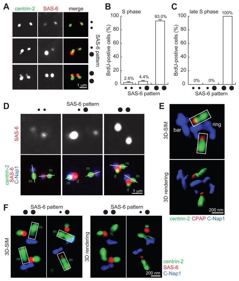
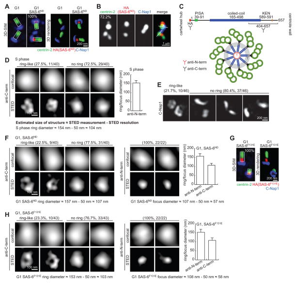
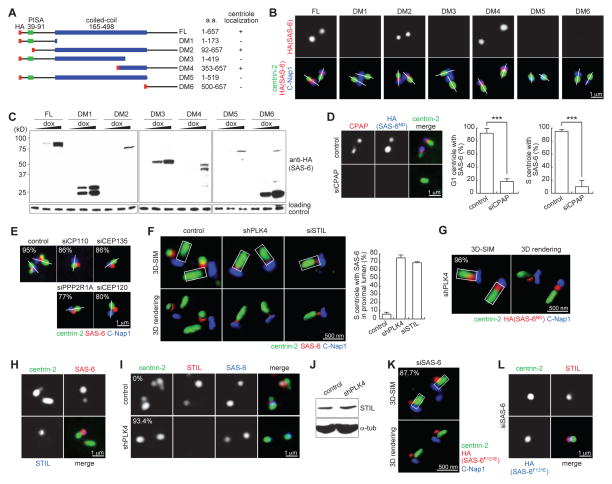
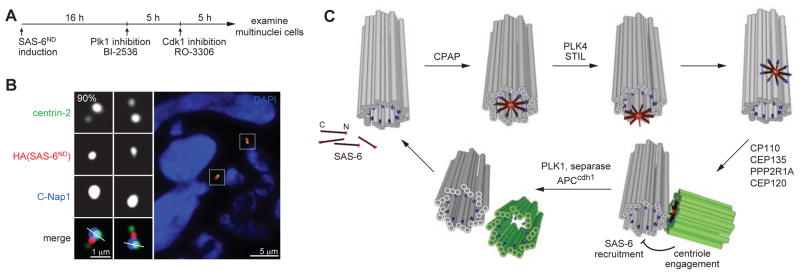
Centrioles are 9-fold symmetric structures duplicating once per cell cycle. Duplication involves self-oligomerization of the centriolar protein SAS-6, but how the 9-fold symmetry is invariantly established remains unclear. Here, we found that SAS-6 assembly can be shaped by preexisting (or mother) centrioles. During S phase, SAS-6 molecules are first recruited to the proximal lumen of the mother centriole, adopting a cartwheel-like organization through interactions with the luminal wall, rather than via their self-oligomerization activity. The removal or release of luminal SAS-6 requires Plk4 and the cartwheel protein STIL. Abolishing either the recruitment or the removal of luminal SAS-6 hinders SAS-6 (or centriole) assembly at the outside wall of mother centrioles. After duplication, the lumen of engaged mother centrioles becomes inaccessible to SAS-6, correlating with a block for reduplication. These results lead to a proposed model that centrioles may duplicate via a template-based process to preserve their geometry and copy number.


Figures

Figure 1
Figure 1. SAS-6 is transiently recruited to the proximal lumen of mother centrioles in early S-phase
(A) U2OS cells in S phase were processed for immunofluorescence to visualize centriole (centrin-2) and SAS-6. (B) Quantification of cells with weak SAS-6 foci in S phase. Error bars, standard deviation. n>80, N=3. (C) Quantification of weak SAS-6 foci in late S phase. n>100, N=3. (D) Weak SAS-6 localizes to the proximal end of mother centrioles. Linear alignment of centrin-2 (distal-end component) and C-Nap1 (proximal end) is marked by a white line. Here, rabbit antibodies recognizing the C-terminus of C-Nap1 were used with a mouse anti-SAS-6 antibody. m, mother; d, daughter. (E) 3D-SIM image of luminal components CPAP and centrin-2 in comparison to non-luminal component C-Nap1. Here, a mouse antibody recognizing the N-terminus of C-Nap1 was used with a rabbit anti-CPAP antibody. Imaris 3D rendering is presented as indicated. (F) 3D-SIM images of SAS-6, C-Nap1, and centrin in S-phase centrioles stained with the antibodies indicated. m, mother. All S-phase cells in (A), (B), (C), (D) and (F) were identified by BrdU labeling (see Experimental Procedures), not shown.
Figure 2
Figure 2. G1 centrioles can recruit and organize SAS-6 into an ordered structure similar to the cartwheel
(A) RPE-1 cells arrested in G1 for 48 hours followed by expression of HA-tagged SAS-6ND were imaged by 3D-SIM. n>45, N=3. (B) G1-arrested RPE-1 cells expressing SAS-6ND as described in (A) were released into S phase by serum reintroduction and stained with the indicated antibodies. S-phase cells were identified by BrdU labeling, not shown. n>21, N=3. (C) Schematic diagram of domain organization of SAS-6 and the epitopes recognized by the antibodies used in STED analyses. (D) Representative STED images of SAS-6 staining with anti-C-term antibody on normal S-phase centrioles were shown. Graph presents STED measurements of ring/focus diameter by averaging the long and short axes. Actual size of the structure is estimated by subtracting STED resolution from STED measurements as indicated. Error bar, standard deviation. n=40. (E) Frequency of observing C-Nap1 ring under 3D-SIM. n=46. (F) STED imaging of SAS-6ND stained with anti-C-term and anti-N-term antibodies. Actual size of the structure is estimated as indicated. Error bars, standard deviation. n as indicated. (G) SAS-6F131E localizes to the proximal lumen of G1 centrioles. 3D-SIM image of G1-arrested RPE-1 cells inducibly expressing HA-tagged SAS-6F131E as described in (A). n>39, N=3. (H) STED imaging of SAS-6F131E stained with anti-C-term and anti-N-term antibodies. Actual size of the structure is estimated as indicated. Error bars, standard deviation. n as indicated.
Figure 3
Figure 3. Molecular requirements for SAS-6 recruitment to and release from centriole lumen
(A) Collections of various SAS-6 deletion mutants tagged with HA (red). Centriolar localizations of these mutants are summarized. (B and C) Localization and expression of SAS-6 mutants. Indicated SAS-6 mutants were inducibly expressed in G1-arrested RPE-1 cells, and their luminal localizations were examined. (D) HeLa cells depleted of CPAP (two sequential RNAi for 6 days) were induced to express SAS-6ND. Centrioles in G1 or S-phase cells were examined for SAS-6 localization as indicated. Similar results were obtained with three independent siRNA oligos (see Experimental Procedures). Error bars, standard deviation. n>38, N=3. The significance (two-tailed t-test) is indicated, *** P < 0.0001. (E) U2OS cells depleted of the indicated protein were examined for the localization of the endogenous SAS-6 during S phase. n>31, N=2. (F) U2OS cells depleted of PLK4 or STIL were examined for the localization of the endogenous SAS-6 during S phase by 3D-SIM. Error bars, standard deviation. n>26, N=3. (G) U2OS cells depleted of PLK4 were induced to express SAS-6ND. The duplication status and localization of the elevated SAS-6ND were examined and quantified in S-phase cells by 3D-SIM with indicated antibodies. Note that duplication was not rescued, and S-phase cells containing unduplicated centrioles (centrin singlet) were counted. n>25, N=3. (H) Wild-type, early S-phase U2OS cells containing one bright and one weak SAS-6 foci were examined for the localization of STIL. Note that STIL only associates with the bright SAS-6 focus. (I) STIL localization in control or PLK4-depleted U2OS cells expressing SAS-6ND was examined. n>29, N=3. (J) Immunoblots showing STIL level is unaffected by PLK4 knockdown. (K and L) SAS-6F131E was inducibly expressed in U2OS cells depleted of the endogenous SAS-6. Localizations of SAS-6F131E (K) and STIL (L) were then examined in S-phase cells by 3D-SIM and regular microscopy, respectively as indicated. n>24, N=3. S-phase cells in (D), (E), (F), (G), (I), (K) and (L) were identified by BrdU labeling, not shown. See also Figure S1.
Figure 4
Figure 4. Engaged centrioles block luminal recruitment of SAS-6
(A) Schematic outlining the generation of G1 cells with engaged centrioles as described previously (Tsou et al., 2009; Wang et al., 2011). SAS-6ND expression was induced in proliferating cells for 16 hours before adding BI-2536 to inhibit Plk1. Mitotic arrested cells were forced to exit mitosis 3 hours after Plk1 inhibition by Cdk1 inhibitor RO-3306 for 5 hours. Multinuclei cells, which were cells that had gone through mitosis in the absence of Plk1 activity, were examined for the centriolar localization of SAS-6 with indicated antibodies. n>26, N=3. (B) SAS-6 recruitment to mother centriole lumen is inhibited by engaged centrioles. Immunofluorescence image of a G1 cell containing engaged centrioles with indicated antibodies. (C) Model for a template-based mechanism of centriole duplication. SAS-6 dimers are individually recruited to the proximal lumen of mother centrioles, where they are organized by the surrounding geometry to undergo 9-fold symmetrical assembly. The assembled SAS-6 oligomer is then released from the lumen and forms the cartwheel that drives daughter centriole formation. Centriole engagement blocks re-recruitment of SAS-6 to the mother centriole lumen and thus prevents centriole re-duplication. Centriole disengagement and cartwheel removal at the end of mitosis allow the centrioles to be used as a template for their own biogenesis. Mother centriole (grey), daughter centriole (green), CPAP (blue), SAS-6 (red).

Comment in

References

    1. Anderson RG, Brenner RM. The formation of basal bodies (centrioles) in the Rhesus monkey oviduct. The Journal of cell biology. 1971;50:10–34. - PMC - PubMed
    1. Arquint C, Nigg EA. STIL microcephaly mutations interfere with APC/C-mediated degradation and cause centriole amplification. Current biology : CB. 2014;24:351–360. - PubMed
    1. Arquint C, Sonnen KF, Stierhof YD, Nigg EA. Cell-cycle-regulated expression of STIL controls centriole number in human cells. Journal of cell science. 2012;125:1342–1352. - PubMed
    1. Azimzadeh J, Wong ML, Downhour DM, Sanchez Alvarado A, Marshall WF. Centrosome loss in the evolution of planarians. Science. 2012;335:461–463. - PMC - PubMed
    1. Cottee MA, Muschalik N, Wong YL, Johnson CM, Johnson S, Andreeva A, Oegema K, Lea SM, Raff JW, van Breugel M. Crystal structures of the CPAP/STIL complex reveal its role in centriole assembly and human microcephaly. eLife. 2013;2:e01071. - PMC - PubMed

Publication types

LinkOut - more resources