Review
. 2014 Aug:258:5-16.
doi: 10.1016/j.expneurol.2014.01.001.

Pattern recognition receptors and central nervous system repair

Affiliations
Review

Pattern recognition receptors and central nervous system repair

Kristina A Kigerl et al. Exp Neurol. 2014 Aug.

Abstract

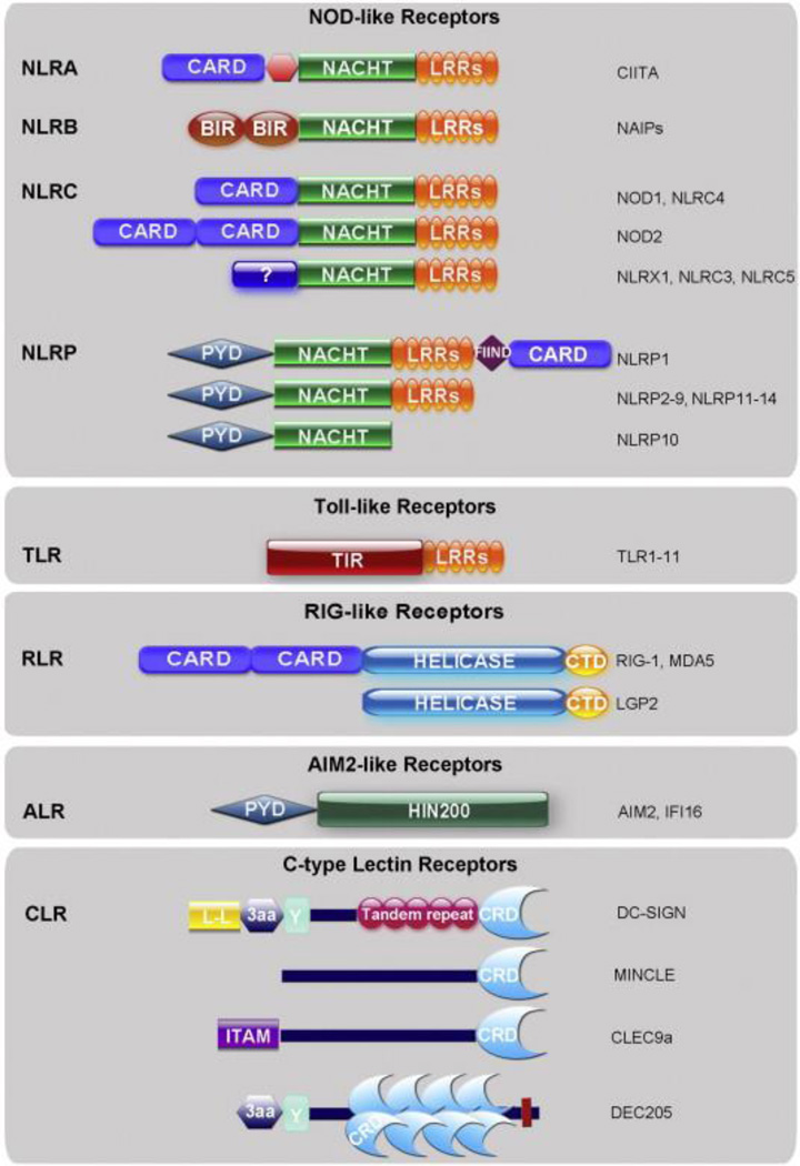
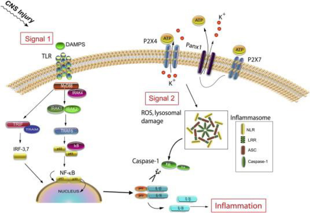
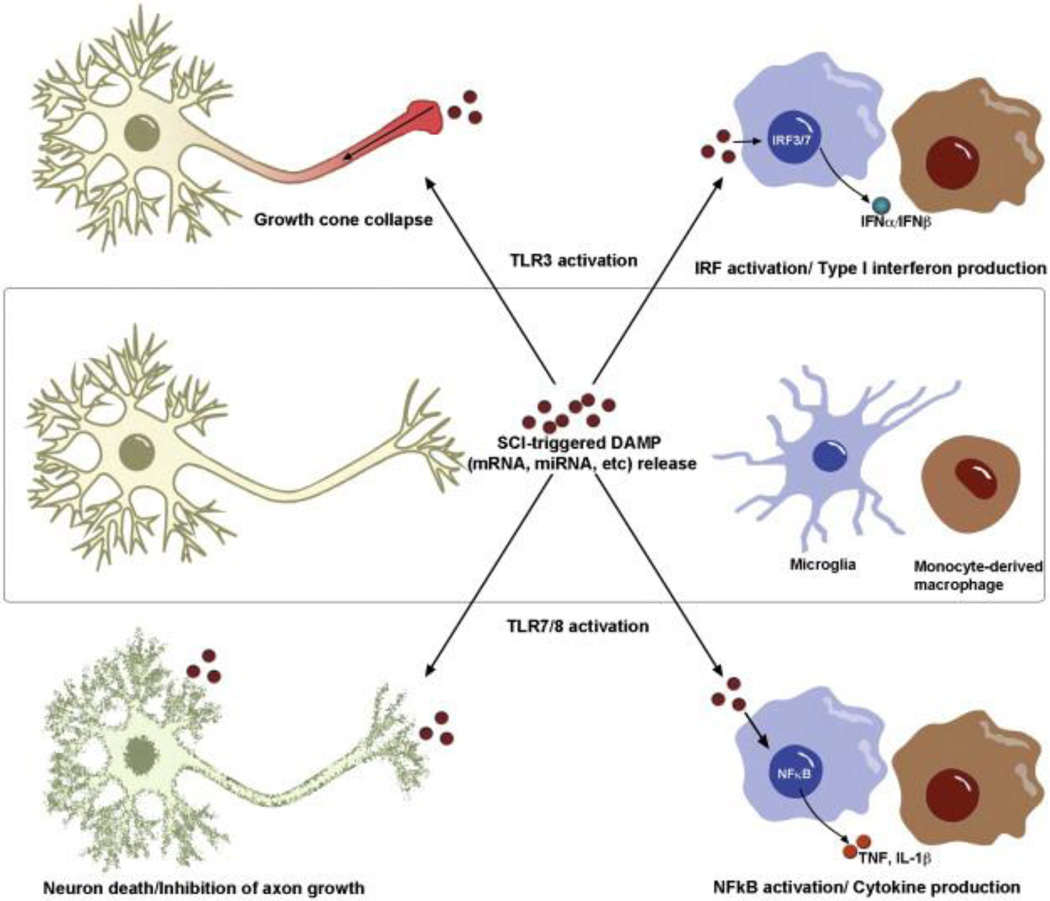
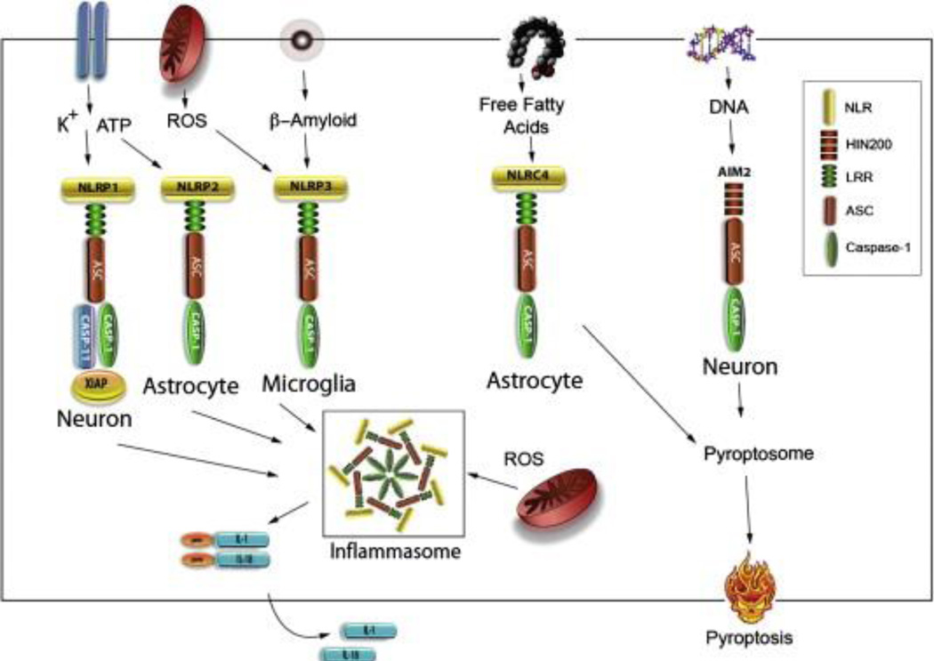
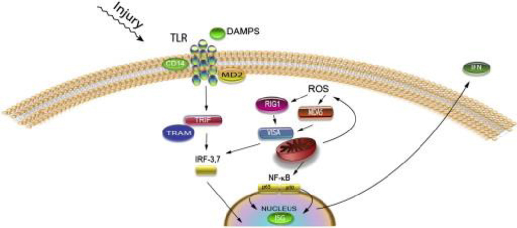
Pattern recognition receptors (PRRs) are part of the innate immune response and were originally discovered for their role in recognizing pathogens by ligating specific pathogen associated molecular patterns (PAMPs) expressed by microbes. Now the role of PRRs in sterile inflammation is also appreciated, responding to endogenous stimuli referred to as "damage associated molecular patterns" (DAMPs) instead of PAMPs. The main families of PRRs include Toll-like receptors (TLRs), Nod-like receptors (NLRs), RIG-like receptors (RLRs), AIM2-like receptors (ALRs), and C-type lectin receptors. Broad expression of these PRRs in the CNS and the release of DAMPs in and around sites of injury suggest an important role for these receptor families in mediating post-injury inflammation. Considerable data now show that PRRs are among the first responders to CNS injury and activation of these receptors on microglia, neurons, and astrocytes triggers an innate immune response in the brain and spinal cord. Here we discuss how the various PRR families are activated and can influence injury and repair processes following CNS injury.

Keywords: Inflammasome; NOD-like receptors; Neuroinflammation; Pattern recognition receptors; Spinal cord injury; Toll-like receptors.


Figures

Fig. 1
Fig. 1
Structural domains of pattern recognition families. The major structural domains of TLRs, NLRs, RLRs, ALRs, and CLRs are depicted above. Note that the NLR family is divided into 4 subfamilies: NLRA, NLRB, NLRC and NLRP. CLRs comprise a large family of receptors, thus only those discussed in this review are shown above. Abbreviations: CARD (caspase recruitment domain), LRR (leucine rich repeat), BIR (baculovirus inhibition of apoptosis protein repeat), PYD (pyrin domain), FIIND (function to find domain), TIR (Toll-IL-1 receptor domain), helicase (DExD/H box helicase domain), CTD (carboxy terminal domain), HIN200 (hematopoietic interferon-inducible nuclear antigens with 200 amino acid repeats), L-L (di-leucine motif), 3aa (triad of acidic amino acids), Y (tyrosine-based motif), CRD (carbohydrate recognition domain), ITAM (immunoreceptor tyrosine-based activation motif).
Fig. 2
Fig. 2
Two-signal model of innate immunity. TLRs and NLRs cooperate to orchestrate the innate immune response to injury. Activation of TLRs (via DAMPs released from CNS injury) leads to nuclear translocation of NFκB and transcription of pro-inflammatory cytokines, such as pro-IL-1β. Activation of NLRs (via a second signal) triggers inflammasome formation, caspase-1 activation, and cleavage of pro-IL-1β into its active form.
Fig. 3
Fig. 3
Activation of “RNA-sensing” TLRs on neurons and microglia triggers distinct effects. Signaling through TLR3 on neurons triggers rapid collapse of growth cones through a non-canonical signaling pathway, while the same ligand–receptor interaction elicits synthesis/release of type I interferons from activated microglia and monocyte-derived macrophages. TLR7/8 activation leads to neuron death and inhibition of axon outgrowth through undefined mechanisms; however, TLR7/8 activation of CNS macrophages triggers canonical TLR signaling pathways leading to NFκB activation and inflammatory cytokine production.
Fig. 4
Fig. 4
Diverse ligands elicit cell-specific inflammasome activation in CNS. High extracellular potassium is required for NLRP1 inflammasome activation in neurons and astrocytes. High concentrations of ATP in the extracellular space stimulate both the NLRP1 and the NLRP2 inflammasome in neurons and astrocytes. The NLRP3 inflammasome is activated by β-amyloid and by ROS. Signals such as ROS appear to be typical activators of all inflammasomes, but thus far has only been shown for NLRP3 inflammasome signaling. NLRC4 inflammasome activation is activated by free fatty acids in astrocytes, and DNA stimulates AIM2 inflammasome activation. Canonical inflammasomes including NLRP1, NLRP3, AIM2 and NLRC4 recruit caspase-1 through interactions with ASC causing pro-IL-1β and pro-IL18 to be cleaved into their active forms IL-1β and IL18. The NRLC4 and AIM2 inflammasome have been involved in the caspase-1 mediated cell death process of pyroptosis by formation of the pyroptosome. NLR: NOD-like receptors, HIN200: hemopoietic IFN-inducible nuclear, LRR: leucine rich repeat, ASC: apoptosis-associated speck-like protein containing a CARD.
Fig. 5
Fig. 5
Production of type I IFN by TLR and RLR signaling: DAMPS binding to TLR signal through TRIF. TRIF recruits TRAF3 (not shown) resulting in phosphorylation of IRF3 or 7, thus activating interferon stimulated genes and type I IFNs such as IFNα and β. ROS activate RLR signaling either through RIG1 or MDA5. Either protein binds to VISA and is recruited to the mitochondrion, resulting in phosphorylation of IRF3 or 7 or activation of NF-κB, resulting in production of interferon stimulated gene proteins and the type I IFNs (IFNα and β).

References

    1. Abulafia DP, de Rivero Vaccari JP, Lozano JD, Lotocki G, Keane RW, Dietrich WD. Inhibition of the inflammasome complex reduces the inflammatory response after thromboembolic stroke in mice. J. Cereb. Blood Flow Metab. 2009;29:534–544. - PubMed
    1. Adamczak SE. Molecular Recognition of DNA by the AIM2 Inflammasome Induces Neuronal Pyroptosis: Implications in Infection and Host Tissue Damage (Open Access Dissertations) 2012
    1. Adamczak S, Dale G, de Rivero Vaccari JP, Bullock MR, Dietrich WD, Keane RW. Inflammasome proteins in cerebrospinal fluid of brain-injured patients as biomarkers of functional outcome: clinical article. J. Neurosurg. 2012;117:1119–1125. - PMC - PubMed
    1. Ahmad F, Wang MY, Levi AD. Hypothermia for acute spinal cord injury — a review. World Neurosurg. 2013 - PubMed
    1. Anderson KV, Bokla L, Nusslein-Volhard C. Establishment of dorsal–ventral polarity in the Drosophila embryo: the induction of polarity by the Toll gene product. Cell. 1985a;42:1791–798. (Epub ahead of print) - PubMed

Publication types

MeSH terms

Substances