. 2014 Aug 7;55(3):495-504.
doi: 10.1016/j.molcel.2014.06.009. Epub 2014 Jul 10.

Mechanism of DNA methylation-directed histone methylation by KRYPTONITE

Affiliations

Mechanism of DNA methylation-directed histone methylation by KRYPTONITE

Jiamu Du et al. Mol Cell. .

Abstract

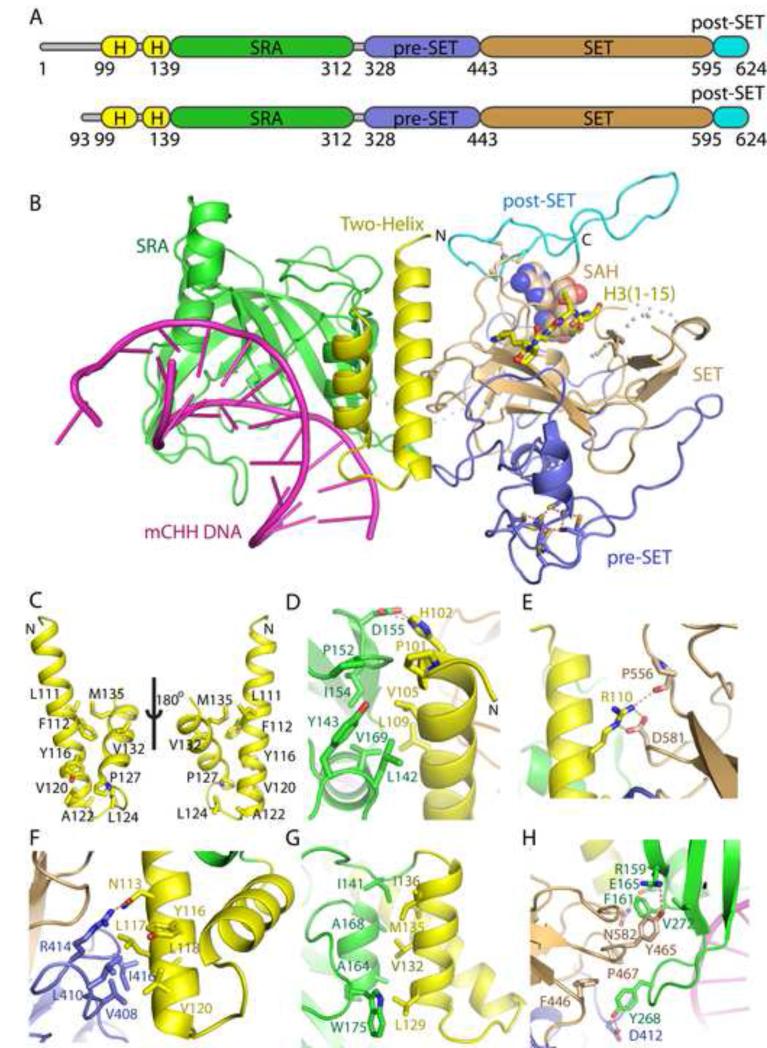
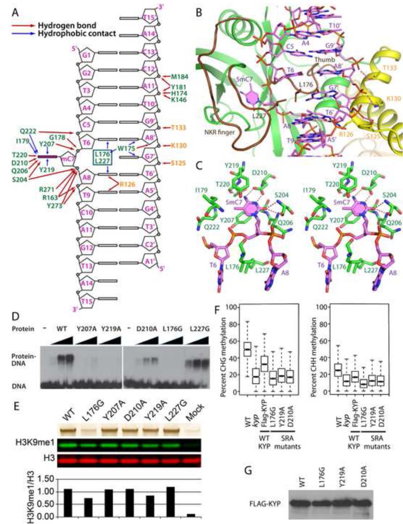
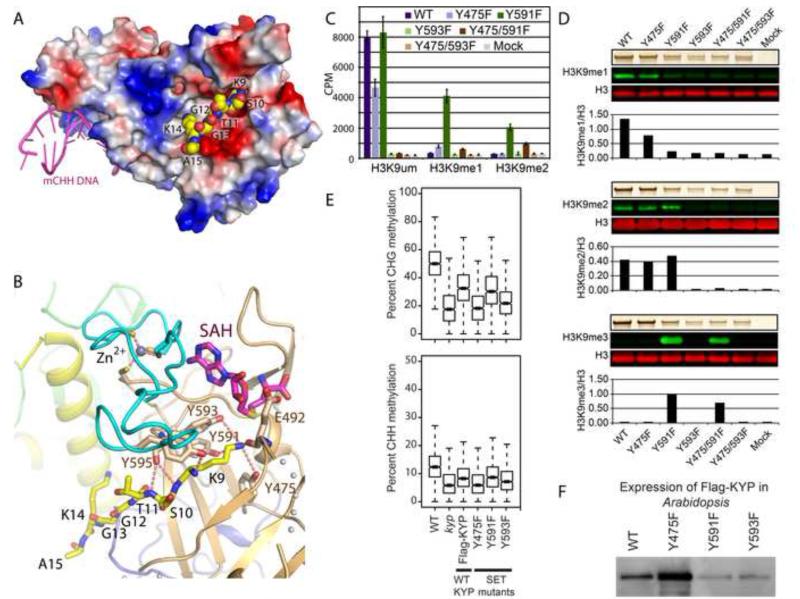
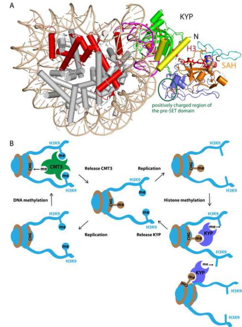
In Arabidopsis, CHG DNA methylation is controlled by the H3K9 methylation mark through a self-reinforcing loop between DNA methyltransferase CHROMOMETHYLASE3 (CMT3) and H3K9 histone methyltransferase KRYPTONITE/SUVH4 (KYP). We report on the structure of KYP in complex with methylated DNA, substrate H3 peptide, and cofactor SAH, thereby defining the spatial positioning of the SRA domain relative to the SET domain. The methylated DNA is bound by the SRA domain with the 5mC flipped out of the DNA, while the H3(1-15) peptide substrate binds between the SET and post-SET domains, with the ε-ammonium of K9 positioned adjacent to bound SAH. These structural insights, complemented by functional data on key mutants of residues lining the 5mC and H3K9-binding pockets within KYP, establish how methylated DNA recruits KYP to the histone substrate. Together, the structures of KYP and previously reported CMT3 complexes provide insights into molecular mechanisms linking DNA and histone methylation.


Figures

Figure 1
Figure 1. Overall Structure of KYP in Complex with mCHH DNA, SAH and H3(1-15) Peptide
(A) Color-coded domain architecture of full length KYP and KYP(93-624) construct used in this study. (B) Ribbon representation of the overall structure of KYP in complex with mCHH DNA, SAH and H3(1-15) peptide. The N-terminal anti-parallel two-helix alignment, SRA, pre-SET, SET and post-SET domains of KYP are color-coded in yellow, green, blue, orange, and cyan, respectively. The mCHH DNA, the SAH cofactor, and the H3 peptide are shown in magenta ribbon, space filling, and stick model, respectively. Some disordered loops, which were not built in the final model, are shown as dashed lines. The Zn3Cys9 triangular zinc cluster in the pre-SET domain is highlighted with ball and stick model. (C) The hydrophobic interactions within the anti-parallel two-helix alignment shown in two views by a 180° rotation. The interacting residues are highlighted in a stick representation. (D) The N-terminal part of the first helix forms extensive hydrophobic and hydrogen bonding interactions with the SRA domain. The interacting residues are shown in stick representation and hydrogen bonds are shown by dashed red line. (E) The middle part of the first helix forms hydrogen bonding as well as salt bridge interactions with the SET domain. (F) The C-terminal part of the first helix forms hydrophobic interactions and hydrogen bonding interaction with the pre-SET domain. (G) One side of the short helix has several hydrophobic residues that form extensive hydrophobic interactions with the SRA domain. (H) The SRA domain forms both hydrophobic and hydrogen bonding interactions with the pre-SET and SET domains. See also Figure S1.
Figure 2
Figure 2. Recognition of mCHH DNA by KYP
(A) Schematic representation of the interactions between KYP and DNA. Hydrogen bonds are shown by red arrows and hydrophobic contacts by blue arrows. The SRA domain residues and the two-helix alignment residues are colored in green and yellow, respectively. (B) The SRA pocket accommodating the flipped-out 5mC and the two-helix alignment residues interacting with the DNA. The 5mC base is highlighted by a solid magenta hexagon. The thumb- and NKR finger-loops are colored in brown with the Leu176 and Leu227 highlighted in a stick representation. The hydrogen bonds between the two-helix alignment residues and mCHH DNA are shown in dashed red lines. (C) The detailed recognition of the flipped out 5mC base by residues lining the binding pocket within the SRA domain. (D) Electrophoretic mobility-shift assays using a mCHH double-stranded DNA and increasing levels (50, 100, and 200 ng) of the indicated protein. Similar results were observed using mCHG substrate (data not shown). (E) In vitro methylation of H3 by KYP SRA domain mutants. KYP protein (upper panel, silver-stained) was incubated with S-adenosyl methionine (SAM) and recombinant H3. Histone methyltransferase activity was tested by quantitative western blot using primary antibodies against H3K9me1 and H3 and infrared secondary antibodies (green: 800 nm, red: 680 nm). (F) Boxplots of CHG and CHH context DNA methylation at a subset of kyp CHG hypomethylated DMRs that show complementation upon transformation with a wild-type KYP construct. All the whiskers on the box plots represent plus/minus 1.5x iqr (inter quartile range). (G) Western blot analysis of lines used in the complementation studies showing expression levels comparable to the Flag-KYP/kyp (WT). See also Figures S2, S3 and Table S1.
Figure 3
Figure 3. Recognition of the Substrate H3 Peptide by the SET Domains of KYP
(A) An electrostatics surface representation of KYP. The bound H3 peptide, in a space filling representation, is inserted into a negatively-charged binding channel. The mCHH DNA in magenta cartoon representation aligns along the opposite side of KYP. (B) The intermolecular interactions amongst SAH, peptide and KYP in the complex. The peptide fits into a narrow channel between the SET (orange) and post-SET (cyan) domains, with the H3K9 inserting into a narrow deep pocket, where it is stabilized through formation of extensive intermolecular hydrogen bonding interactions. The zinc-binding motif, which stabilizes the fold of the post-SET domain, is highlighted with ball and stick representation. (C) Methyltransferase activity of KYP WT and SET domain mutants. Radioactivity (CPM) of H3 peptides was measured after incubation of unmethylated (H3K9um), monomethylated (H3K9me1) or dimethylated (H3K9me2) substrate with KYP protein and tritiated SAM (n = 3, ± S.D.). (D) Differential in vitro methylation of H3 by KYP SET domain mutants. KYP protein (upper panels, silver-stained) was incubated with SAM and recombinant H3. Histone methyltransferase activity was tested by quantitative western blots using primary antibodies against H3 and H3K9me1, H3K9me2 or H3K9me3 and infrared secondary antibodies (green: 800 nm, red: 680 nm). (E) Boxplots of CHG and CHH context DNA methylation at a subset of kyp CHG hypomethylated DMRs that show complementation upon transformation with a wild-type KYP construct (see Supplemental Methods). (F) Western blot analysis of lines used in the complementation studies showing expression levels comparable to the Flag-KYP/kyp (WT).
Figure 4
Figure 4. A Working Model for the Epigenetics Mechanism Controlling H3K9me by KYP
(A) Modeling of KYP on the nucleosomal DNA indicates that KYP can be bound to methylated nucleosomal DNA and further methylate the H3 tail of the same nucleosome. The KYP is colored as Figure 1B. The H3 is highlighted in red. The nucleosomal DNA and other histone proteins are colored in wheat and silver, respectively. The flipped out 5mC is highlighted in space filling model to indicate its position. A green circle marks a positively charged region within the pre-SET domain that is adjacent the nucleosomal DNA, indicating plausible interaction between them. (B) A schematic model of CMT3 and KYP controlled self-reinforcing feedback loop between mCHG and H3K9me.

References

    1. Adams PD, Afonine PV, Bunkoczi G, Chen VB, Davis IW, Echols N, Headd JJ, Hung LW, Kapral GJ, Grosse-Kunstleve RW, et al. PHENIX: a comprehensive Python-based system for macromolecular structure solution. Acta Crystallogr D Biol Crystallogr. 2010;66:213–221. - PMC - PubMed
    1. Arita K, Ariyoshi M, Tochio H, Nakamura Y, Shirakawa M. Recognition of hemi-methylated DNA by the SRA protein UHRF1 by a base-flipping mechanism. Nature. 2008;455:818–821. - PubMed
    1. Avvakumov GV, Walker JR, Xue S, Li Y, Duan S, Bronner C, Arrowsmith CH, Dhe-Paganon S. Structural basis for recognition of hemi-methylated DNA by the SRA domain of human UHRF1. Nature. 2008;455:822–825. - PubMed
    1. Bartee L, Malagnac F, Bender J. Arabidopsis cmt3 chromomethylase mutations block non-CG methylation and silencing of an endogenous gene. Genes Dev. 2001;15:1753–1758. - PMC - PubMed
    1. Cedar H, Bergman Y. Linking DNA methylation and histone modification: patterns and paradigms. Nat Rev Genet. 2009;10:295–304. - PubMed

Publication types

MeSH terms