

Functional Segregation of the Entopallium in Pigeons
- PMID: 25018563
- PMCID: PMC4091902
Functional Segregation of the Entopallium in Pigeons
Abstract
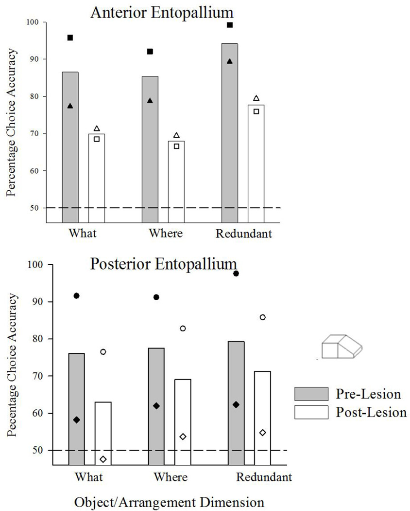
In birds, the entopallium is the primary telencephalic target of the major visual ascending route called the tectofugal pathway. Often functionally compared to the primate geniculo-striate pathway and its subsequent telencephalic (cortical) regions, the latter processes visual information in a parallel fashion in terms of anatomy, physiology, and function. Little is known, however, about the exact mechanism of whether or how information is segregated or integrated in the avian tectofugal pathway including the telencephalon. Testing four pigeons, we examined whether or not color, form, and motion information is selectively processed by different portions of the entopallium. Each learned three distinct visual tasks requiring discrimination of different combinations of color, form and motion cues. After learning and pre-lesion testing, two pigeons received lesions to the anterior portion of the entopallium and two received lesions to the posterior entopallium. During post-lesion testing the pigeons with anterior lesions exhibited significant deficits in those tasks most dependent on color and form discrimination, but showed no deficit on a task that had involved discriminating among forms that were moving. Pigeons with posterior lesions showed a different pattern of deficits, exhibiting significant reductions in discriminating both moving and static forms, but little or no deficits in color discrimination. These divergent profiles of effects for each lesion suggest there is a functional segregation of visual information processing in the pigeon telencephalon. This indicates a convergence between birds and primates regarding the parallel processing and separation of information within their phylogenetically distinct major visual pathways.
Keywords: birds; lesion; visual system.
Figures


References
-
- Asen YL, Cook RG. Discrimination and categorization of actions by pigeons. Psychol. Sci. 2012;23:617–624. - PubMed
-
- Cook RG. Acquisition and transfer of visual texture discriminations by pigeons. J. Exp. Psychol. Anim. B. 1992a;18:341–353.
-
- Cook RG. Dimensional organization and texture discrimination in pigeons. J. Exp. Psychol. Anim. B. 1992b;18:351–363.
-
- Cook RG. The visual perception and processing of textures by pigeons. In: Honig WK, Fetterman JG, editors. Cognitive aspects of stimulus control. New Jersey: Lawrence Erlbaum Associates, Inc.; 1992c. pp. 279–299.
-
- Cook RG. The comparative psychology of avian visual cognition. Curr. Dir.In Psychol. Sci. 2000;9:83–89.
Grants and funding
LinkOut - more resources
Full Text Sources