. 2013 Fall;10(3):e127-e135.
doi: 10.1016/j.ddmod.2012.08.004.

MicroRNAs are potential therapeutic targets in fibrosing kidney disease: lessons from animal models

Affiliations

MicroRNAs are potential therapeutic targets in fibrosing kidney disease: lessons from animal models

Jeremy S Duffield et al. Drug Discov Today Dis Models. 2013 Fall.

Abstract

Chronic disease of the kidneys has reached epidemic proportions in industrialized nations. New therapies are urgently sought. Using a combination of animal models of kidney disease and human biopsy samples, a pattern of dysregulated microRNA expression has emerged which is common to chronic diseases. A number of these dysregulated microRNA have recently been shown to have functional consequences for the disease process and therefore may be potential therapeutic targets. We highlight microRNA-21, the most comprehensively studied microRNA in the kidney so far. MicroRNA-21 is expressed widely in healthy kidney but studies from knockout mice indicate it is largely inert. Although microRNA-21 is upregulated in many cell compartments including leukocytes, epithelial cells and myofibroblasts, the inert microRNA-21 also appears to become activated, by unclear mechanisms. Mice lacking microRNA-21 are protected from kidney injury and fibrosis in several distinct models of kidney disease, and systemically administered oligonucleotides that specifically bind to the active site in microRNA-21, inhibiting its function, recapitulate the genetic deletion of microRNA-21, suggesting inhibitory oligonucleotides may have therapeutic potential. Recent studies of microRNA-21 targets in kidney indicate that it normally functions to silence metabolic pathways including fatty acid metabolism and pathways that prevent Reactive Oxygen Species generation in peroxisomes and mitochondria in epithelial cells and myofibroblasts. Targeting specific pathogenic microRNAs in a specific manner is feasible in vivo and may be a new therapeutic target in disease of the kidney.

Keywords: PPARα; ROS; chronic allograft dysfunction; chronic kidney disease; fibrosis; microRNA.


Conflict of interest statement

Conflict of Interest

JSD serves on the Scientific Advistoy Board of Regulus therpeutics and has a Research Grant sponsored by Regulus Therapeutics.

Figures
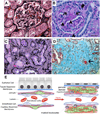
Figure 1
Figure 1. Characteristic manifestations and model of disease mechanisms of chronic kidney disease in glomeruli and interstitium of human kidney cortex
(A) Normal human glomerulus and surrounding tubules and peritubular capillaries (PTCs) filled with erythrocytes (* placed above examples of PTCs) stained with Silver methenamine combined with PAS (Jones) stain which highlights collagens. Arteriole (a) is shown. Note back to back tubules with cuboidal or columnar epithelium. (B) Sclerotic glomerulus showing wedge shaped sclerotic region showing dense pink material on PAS stained section (arrowhead) and rather acellular weaker pink stained material more peripherally (arrow) and obliteration of capillary loops. Also note the sclerotic region is fused to Bowman’s capsule where there is local destruction of the basement membrane and periglomerular fibrosis (thick arrow). At the lower pole note a combination of increased cellularity and fibrosis in the mesangium, and basement membrane thickening in glomerular loops. (C) Jones stained image of cortex from diabetic nephropathy, showing injured tubules (tubule atrophy and tubule cell vacuolization, apoptotic cells, arrow), marked reduction in capillary density (* placed adjacent to examples of PTCs), expansion of the interstitial space with fibrotic material (fine black stain), and an increase in inflammatory cells. Note also thickening of the tubule basement membrane (black) (D) Trichrome stained image of kidney cortex from ischemic kidney disease showing marked expansion of interstitial fibrosis (cyan color) which has overtaken all of the tubules. The fibrosis is cellular showing inflammatory cells and myofibroblasts. The remaining tubules all show tubular atrophy with intraluminal debris. (E) Schema showing cellular mechanisms of CKD development in the kidney cortical interstitium.
Figure 2
Figure 2. Images of kidney disease after in mice that lack miR-21 compare with WT mice following kidney injury
Sirius red stained low power images showing red stained interstitial fibrosis following kidney injury, and medium power images showing PAS stain of kidney cortex after injury. Not reduced fibrosis in kidneys lacking miR-21. Note also that kidney epithelial cells are more injured in WT showing increased flattening and loss of typical purple brush border, whereas these features are more preserved in miR21−/− kidneys.
Figure 3
Figure 3. Schema showing major gene products and pathways silenced by miR-21 in animal models of kidney disease
MiR-21 silences the transcriptional regulator PPARα and many of the downstream enzymes in fatty acid metabolism that are also regulated by PPARα, including transporters and enzymes (shown in grey) of the β-oxidation metabolic pathway of fatty acids that occurs in peroxisomes and mitochondria. The consequence of miR-21 activity is to reduce metabolism of fatty acids. In addition miR-21 increases reactive oxygen species and toxin formation/accumulation. Firstly by suppressing peroxisome formation and activity, the metabolism of H2O2 is retarded by miR-21 and secondly miR-21 silences genes that inhibit ROS generation in mitochondria, including MPV17-like which exerts its inhibitory function by binding to the mitochondrial protease HrtA2. In this configuration HrtA2 is also anti-apoptotic.

References

    1. Snyder JJ, et al. Prevalence of CKD in the United States: a sensitivity analysis using the National Health and Nutrition Examination Survey (NHANES) 1999–2004. Am J Kidney Dis. 2009;53(2):218–228. - PMC - PubMed
    1. Johnson RJ, et al. Subtle acquired renal injury as a mechanism of saltsensitive hypertension. N Engl J Med. 2002;346(12):913–923. - PubMed
    1. Friedman SL, et al. Therapy for Fibrotic Diseases: Nearing the Start Line. Science Trans. Med. 2012 in press. - PubMed
    1. Ishii Y, et al. Injury and progressive loss of peritubular capillaries in the development of chronic allograft nephropathy. Kidney Int. 2005;67(1):321–332. - PubMed
    1. Schrimpf C, Duffield JS. Mechanisms of fibrosis: the role of the pericyte. Curr Opin Nephrol Hypertens. 2011;20(3):297–305. - PubMed