. 2014 Jun 26:4:168.
doi: 10.3389/fonc.2014.00168. eCollection 2014.

Aberrant Proliferation of Differentiating Alveolar Cells Induces Hyperplasia in Resting Mammary Glands of SV40-TAg Transgenic Mice

Affiliations

Aberrant Proliferation of Differentiating Alveolar Cells Induces Hyperplasia in Resting Mammary Glands of SV40-TAg Transgenic Mice

Timo Quante et al. Front Oncol. .

Abstract

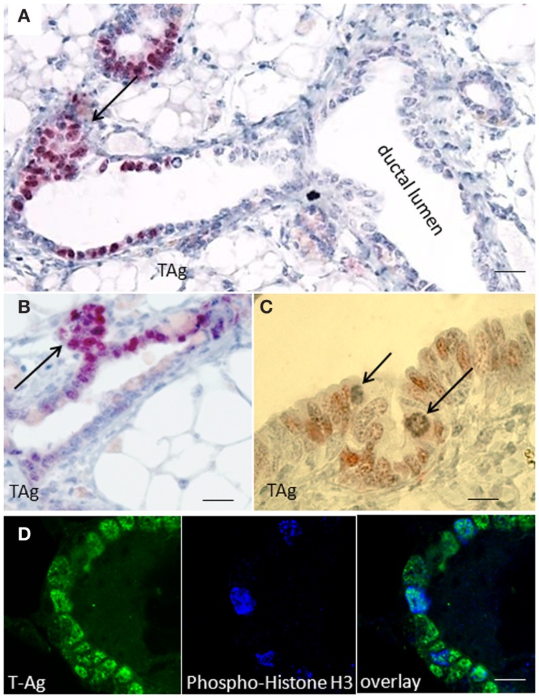
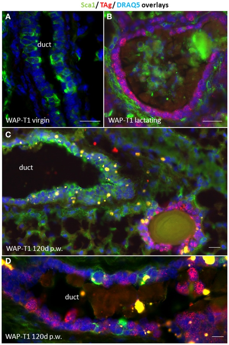
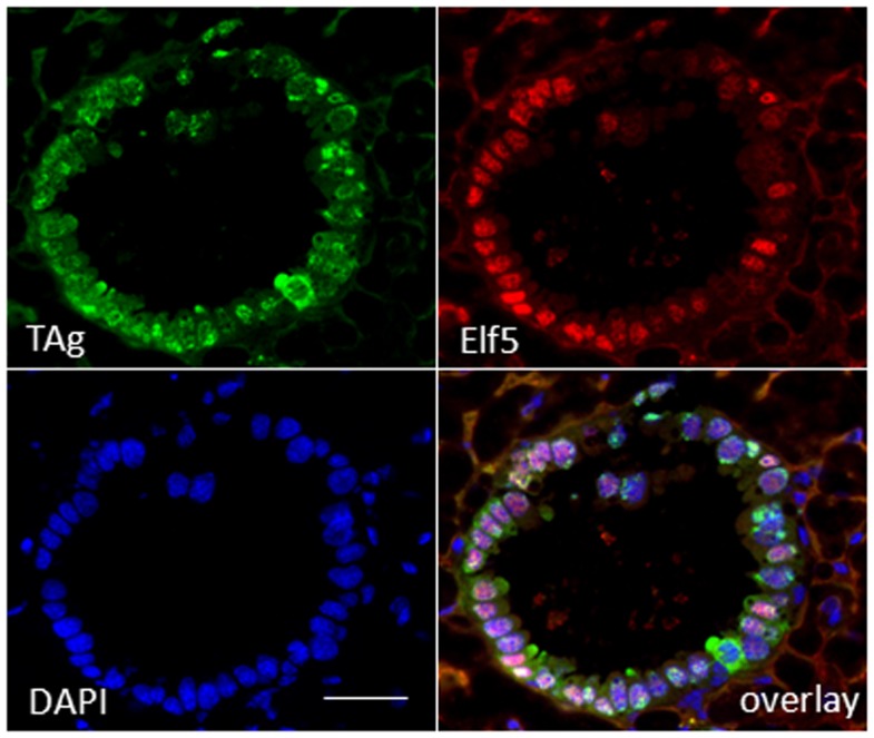
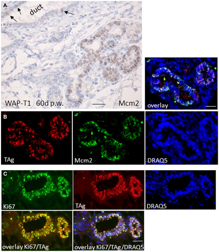
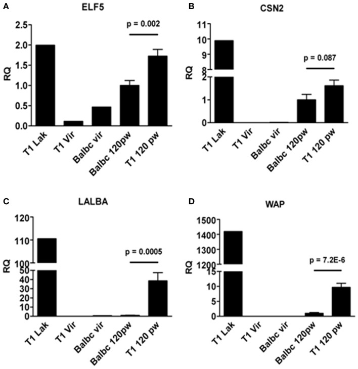
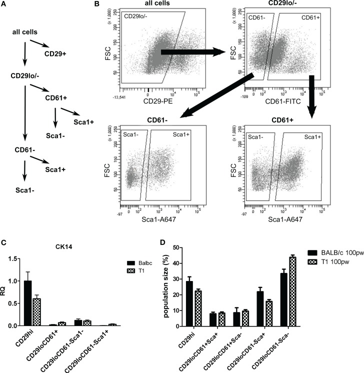
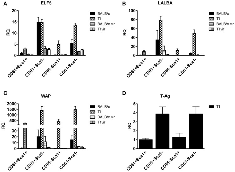
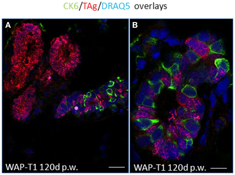
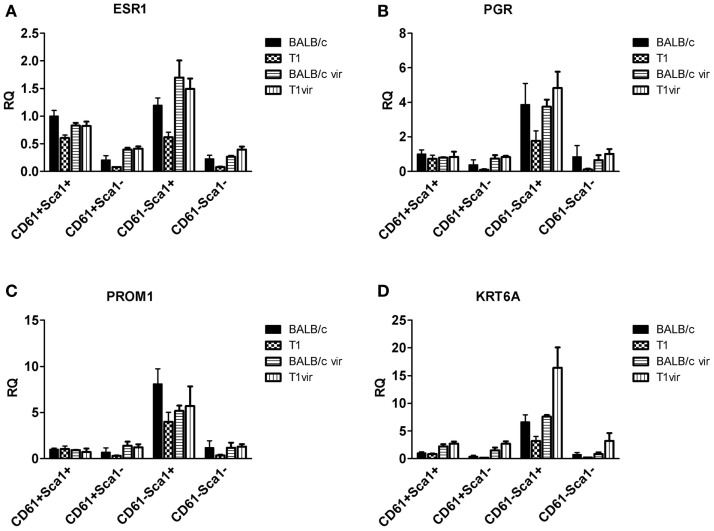
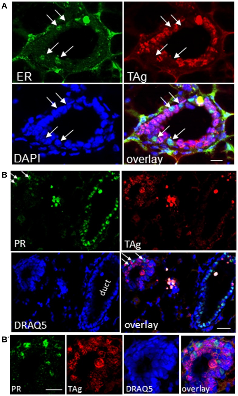
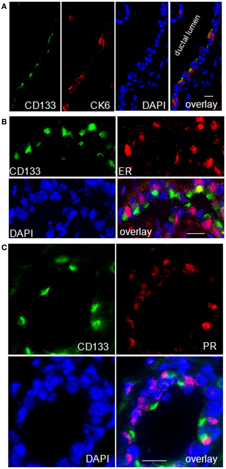
WAP-T1 transgenic mice express SV40-TAg under control of the whey acidic protein (WAP) promoter, which directs activity of this strong viral oncogene to luminal cells of the mammary gland. Resting uniparous WAP-T1 glands develop hyperplasia composed of TAg positive cells prior to appearance of advanced tumor stages. We show that cells in hyperplasia display markers of alveolar differentiation, suggesting that TAg targets differentiating cells of the alveolar compartment. The glands show significant expression of Elf5 and milk genes (Lalba, Csn2, and Wap). TAg expressing cells largely co-stain with antibodies to Elf5, lack the epithelial marker Sca1, and are hormone receptor negative. High expression levels of Elf5 but not of milk genes are also seen in resting glands of normal BALB/c mice. This indicates that expression of Elf5 in resting WAP-T1 glands is not specifically induced by TAg. CK6a positive luminal cells lack TAg. These cells co-express the markers prominin-1, CK6a, and Sca1, and are positive for hormone receptors. These hormone sensitive cells localize to ducts and seem not to be targeted by TAg. Despite reaching an advanced stage in alveolar differentiation, the cells in hyperplasia do not exit the cell cycle. Thus, expression of TAg in conjunction with regular morphogenetic processes of alveologenesis seem to provide the basis for a hormone independent, unscheduled proliferation of differentiating cells in resting glands of WAP-T1 transgenic mice, leading to the formation of hyperplastic lesions.

Keywords: SV40-TAg; alveologenesis; hyperplasia; mammary gland; tumorigenesis.


Figures

Figure 1
Figure 1
SV40-TAg positive epithelial cells cluster at sites of bud formation. (A–C) TAg labeling on sections of paraffin embedded WAP-T1 glands isolated 60 days post-weaning (p.w.); alkaline phosphatase (A,B) or peroxidase (C) conjugated antibodies were used as secondary antibodies; TAg expressing cells cluster at sites of bud formation [arrows in (A,B)]; individual TAg positive epithelial cells show condensing chromatin [arrows in (C)]; (D) IF double labeling with antibodies to TAg (green) and phospho-histone H3 (blue) on cryosections of resting uniparous WAP-T1 glands; phospho-histone H3 staining points to mitotic activity in WAP-T1 hyperplastic lesions. Bars: (A) = 50 μm; (B) = 15 μm; (C,D)  = 10 μm.
Figure 2
Figure 2
TAg is expressed in Sca1 negative luminal epithelial cells. (A,B) IF double labeling with antibodies to Sca1(green) and TAg (red) on cryosections of WAP-T1 glands; staining of DNA with DAPI (blue); (A) luminal epithelia in virgin WAP-T1 glands are TAg negative and reveal a heterogenous Sca1 staining pattern; (B) luminal epithelia in lactating WAP-T1 glands are TAg positive and Sca1 negative; (C,D) TAg positive cells in epithelial compartments of resting uniparous WAP-T1 glands (120 days p.w.) are also Sca1 negative. Bars: (A–C) = 50 μm;(D) = 5 μm.
Figure 3
Figure 3
TAg positive cells in hyperplasia co-stain with antibodies to ELF5. IF double labeling on cryosections of resting uniparous WAP-T1 glands (120 days p.w.) with antibodies to TAg (green) and ELF5 (red); staining of DNA with DAPI; individual TAg positive cells lack ELF staining signal. Bar = 50 μm.
Figure 4
Figure 4
TAg positive luminal epithelial cells co-stain with antibodies to proliferation markers. (A) Immunoperoxidase labeling with Mcm2 antibodies on sections of paraffin embedded resting uniparous WAP-T1 glands (60 days p.w.) shows strong nuclear staining of epithelia in hyperplasia; in ductal epithelia only individual cells stain positively for Mcm2 (arrows); (B,C) IF double labeling on cryosections from resting uniparous WAP-T1 glands (120 days p.w.) shows coincident labeling of TAg (red) and Mcm2 (green) (B), respectively TAg (red) and Ki67 (green) (C); DNA staining with DRAQ5 (blue) Bars: (A,B) = 20 μm.
Figure 5
Figure 5
qRT-PCR analysis of ELF5 and milk gene expression in whole glands. ELF5 (A) and milk genes (CSN2, LALBA, WAP) (B–D) are expressed in resting glands (120 days p.w.) from BALB/c and WAP-T1 mice (n = 10 from five mice); ELF5, (A), LALBA (C), and WAP (D) levels are significantly higher in WAP-T1 than in BALB/c glands; individual glands from lactating WAP-T1 mice (T1 Lak), WAP-T1 virgin mice (T1Vir), and BALB/c virgin mice (Balbc vir) are included for comparison; data presented as mean ± SEM.
Figure 6
Figure 6
Sorting strategy for isolation of luminal epithelial cell populations. After lineage depletion, basal, myoepithelial cells (CD29+) were separated from luminal epithelial cells (CD29lo/−). Luminal epithelial cells were separated into undifferentiated (CD61+) and differentiated (CD61) subpopulations and further separated into Sca1 and Sca1+ subpopulations (A,B). (C) CK14, a marker of myoepithelial cells, is enriched in the basal CD29hi subpopulation (n = 5). (D) Sizes of subpopulations from BALB/c (n = 10) and WAP-T1 (n = 23) mice as percent total cells; data presented as mean ± SEM. Population size of the CD61/Sca1+ population was significantly decreased in WAP-T1 mice compared to BALB/c (15.6 vs. 27.9%, p < 0.05) while CD61/Sca1 population size was significantly increased (54.2 vs. 39.2%, p < 0.05).
Figure 7
Figure 7
qRT-PCR analysis of ELF5, milk gene (LALBA, WAP), and TAg expression in luminal epithelial cell subpopulations from resting (120 days p.w.) glands of uniparous mice (BALB/c, T1) and glands from virgin mice (BALB/c vir, T1 vir). ELF5 (A), LALBA (B), WAP (C), and TAg (D) expression are most prominent in Sca1 negative cells; note equivalent ELF5 levels in undifferentiated (CD61+Sca1) cells of BALB/c and T1, but different levels in differentiated (CD61Sca1) cells from both mouse strains; virgin mice show only basal expression of ELF5 and milk genes (BALB/c n = 4, T1 n = 5, BALB/c vir n = 3, T1 vir n = 3); ata presented as mean ± SEM.
Figure 8
Figure 8
TAg does not target CK6a positive luminal epithelial cells in resting uniparous WAP-T1 glands. IF double labeling with antibodies to CK6a (green) and TAg (red) on cryosections from resting uniparous WAP-T1 glands (120 days p.w.); DNA staining with DRAQ5 (blue); TAg positive lesions lack CK6a positive cells (A); CK6a and TAg mark different luminal epithelial cells in ductal compartments (A,B). Bars: (A) = 30 μm;(B) = 5 μm.
Figure 9
Figure 9
qRT-PCR analysis of hormone receptor, prominin-1, and Ck6a expression in luminal cell subpopulations. Expression of the estrogen receptor (ESR1) (A) and progesterone receptor (PGR) (B) in the CD61Sca1+ ductal cell subpopulation coincides with expression of prominin-1 (PROM1) (C) and CK6a (KRT6A) (D); subpopulations from virgin mice (BALB/c vir n = 3; T1vir n = 3); subpopulations from uniparous mice 120 days p.w. (BALB/c n = 4; T1 n = 5); data presented as mean ± SEM.
Figure 10
Figure 10
TAg positive epithelia in resting uniparous WAP-T1 glands are negative for estrogen receptor (ER) and progesterone receptor (PR). (A–B′): IF double labeling on cryosections from resting uniparous WAP-T1 glands; (A) Double labeling with antibodies to estrogen receptor (ER) (green) and TAg (red); staining of DNA with DAPI (blue); ER positive cells are TAg negative [arrows in (A)]; (B,B′): double labeling with antibodies to progesterone receptor (PR) (green) and TAg (red); DNA staining with DAPI (blue); note the high number of PR positive but TAg negative cells in epithelia of ducts; no coincident labeling of PR [arrows in (B)] and TAg. Bars: (A) = 20 μm;(B,B′) = 50 μm.
Figure 11
Figure 11
CK6a marks CD133 (prominin-1) and hormone receptor positive cells in luminal epithelia of WAP-T1 glands. (A–C) IF double labeling on cryosections of resting uniparous WAP-T1 glands (120 days p.w.); DNA staining with DAPI (blue); (A) Coincident staining of luminal epithelial cells in ducts with antibodies to CD133 (green) and CK6 (red); (B) Coincident labeling of luminal cells with antibodies to CD133 (green) and estrogen receptor (red); (C) Coincident staining of luminal epithelial cells with antibodies to CD133 and progesterone receptor (PR); note the cap-like staining of CD133 at the luminal side of epithelial cells in (A–C). Bars: (A) = 20 μm;(B,C) = 50 μm.

Similar articles

Cited by

References

    1. Medina D. Biological and molecular characteristics of the premalignant mouse mammary gland. Biochim Biophys Acta (2002) 1603(1):1–910.1016/S0304-419X(02)00053-7 - DOI - PubMed
    1. Mastracci TL, Boulos FI, Andrulis IL, Lam WL. Genomics and premalignant breast lesions: clues to the development and progression of lobular breast cancer. Breast Cancer Res (2007) 9(6):215.10.1186/bcr1785 - DOI - PMC - PubMed
    1. Sinn HP, Elsawaf Z, Helmchen B, Aulmann S. Early breast cancer precursor lesions: lessons learned from molecular and clinical studies. Breast Care (Basel) (2010) 5(4):218–2610.1159/000319624 - DOI - PMC - PubMed
    1. Costarelli L, Campagna D, Mauri M, Fortunato L. Intraductal proliferative lesions of the breast-terminology and biology matter: premalignant lesions or preinvasive cancer? Int J Surg Oncol (2012) 2012:501904.10.1155/2012/501904 - DOI - PMC - PubMed
    1. Abdel-Fatah TM, Powe DG, Hodi Z, Reis-Filho JS, Lee AH, Ellis IO. Morphologic and molecular evolutionary pathways of low nuclear grade invasive breast cancers and their putative precursor lesions: further evidence to support the concept of low nuclear grade breast neoplasia family. Am J Surg Pathol (2008) 32(4):513–2310.1097/PAS.0b013e318161d1a5 - DOI - PubMed