

Occurrence of spontaneous periodontal disease in the SAMP1/YitFc murine model of Crohn disease
- PMID: 25019175
- PMCID: PMC4460836
- DOI: 10.1902/jop.2014.140316
Occurrence of spontaneous periodontal disease in the SAMP1/YitFc murine model of Crohn disease
Abstract
Background: Oral involvement is often associated with inflammatory bowel disease (IBD). Recent evidence suggests a high incidence of periodontal disease in patients with Crohn disease (CD). To the best of the authors' knowledge, no animal model of IBD that displays associated periodontal disease was reported previously. The aim of this study is to investigate the occurrence and progression of periodontal disease in SAMP1/YitFc (SAMP) mice that spontaneously develop a CD-like ileitis. In addition, the temporal correlation between the onset and progression of periodontal disease and the onset of ileitis in SAMP mice was studied.
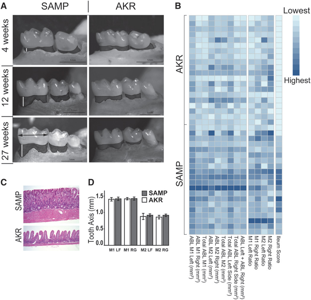
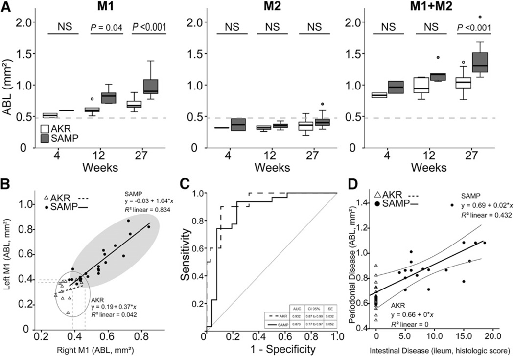
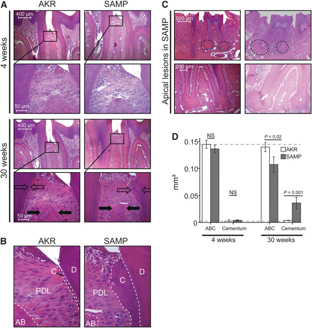
Methods: At different time points, SAMP and parental AKR/J (AKR) control mice were sacrificed, and mandibles were prepared for stereomicroscopy and histology. Terminal ilea were collected for histologic assessment of inflammation score. Periodontal status, i.e., alveolar bone loss (ABL) and alveolar bone crest, was examined by stereomicroscopy and histomorphometry, respectively.
Results: ABL increased in both strains with age. SAMP mice showed greater ABL compared with AKR mice by 12 weeks of age, with maximal differences observed at 27 weeks of age. AKR control mice did not show the same severity of periodontal disease. Interestingly, a strong positive correlation was found between ileitis severity and ABL in SAMP mice, independent of age.
Conclusions: The present results demonstrate the occurrence of periodontal disease in a mouse model of progressive CD-like ileitis. In addition, the severity of periodontitis strongly correlated with the severity of ileitis, independent of age, suggesting that common pathogenic mechanisms, such as abnormal immune response and dysbiosis, may be shared between these two phenotypes.
Keywords: Crohn disease; animal; inflammatory bowel diseases; models; periodontal diseases; periodontitis..
Conflict of interest statement
The authors report no conflicts of interest related to this study.
Figures
Similar articles
-
The Artificial Sweetener Splenda Promotes Gut Proteobacteria, Dysbiosis, and Myeloperoxidase Reactivity in Crohn's Disease-Like Ileitis.Inflamm Bowel Dis. 2018 Apr 23;24(5):1005-1020. doi: 10.1093/ibd/izy060. Inflamm Bowel Dis. 2018. PMID: 29554272 Free PMC article.
-
A novel model of colitis-associated cancer in SAMP1/YitFc mice with Crohn's disease-like ileitis.PLoS One. 2017 Mar 16;12(3):e0174121. doi: 10.1371/journal.pone.0174121. eCollection 2017. PLoS One. 2017. PMID: 28301579 Free PMC article.
-
Death Receptor 3 Signaling Controls the Balance between Regulatory and Effector Lymphocytes in SAMP1/YitFc Mice with Crohn's Disease-Like Ileitis.Front Immunol. 2018 Mar 1;9:362. doi: 10.3389/fimmu.2018.00362. eCollection 2018. Front Immunol. 2018. PMID: 29545797 Free PMC article.
-
SAMP1/YitFc mouse strain: a spontaneous model of Crohn's disease-like ileitis.Inflamm Bowel Dis. 2011 Dec;17(12):2566-84. doi: 10.1002/ibd.21638. Epub 2011 May 6. Inflamm Bowel Dis. 2011. PMID: 21557393 Free PMC article. Review.
-
Pathogenesis of gastritis in ileitis-prone SAMP1/Yit mice.Keio J Med. 2011;60(2):65-8. doi: 10.2302/kjm.60.65. Keio J Med. 2011. PMID: 21720202 Review.
Cited by
-
Identification and Validation of Signature Genes and Potential Therapy Targets of Inflammatory Bowel Disease and Periodontitis.J Inflamm Res. 2023 Sep 28;16:4317-4330. doi: 10.2147/JIR.S426004. eCollection 2023. J Inflamm Res. 2023. PMID: 37795494 Free PMC article.
-
Periodontitis and Inflammatory Bowel Disease: A Review.Cureus. 2024 Feb 20;16(2):e54584. doi: 10.7759/cureus.54584. eCollection 2024 Feb. Cureus. 2024. PMID: 38523972 Free PMC article. Review.
-
Vitamin D Axis in Inflammatory Bowel Diseases: Role, Current Uses and Future Perspectives.Int J Mol Sci. 2017 Nov 7;18(11):2360. doi: 10.3390/ijms18112360. Int J Mol Sci. 2017. PMID: 29112157 Free PMC article. Review.
-
Integrated oral-gut microbiota therapy: a novel perspective on preventing bacterial translocation for systemic disease management.Front Cell Infect Microbiol. 2025 Jul 28;15:1641816. doi: 10.3389/fcimb.2025.1641816. eCollection 2025. Front Cell Infect Microbiol. 2025. PMID: 40792109 Free PMC article. Review.
-
The Potential Association Between Inflammatory Bowel Diseases and Apical Periodontitis: A Systematic Review and Meta-Analysis.Eur Endod J. 2024 Jan 1;9(1):8-17. doi: 10.14744/eej.2023.74507. Epub 2023 Nov 16. Eur Endod J. 2024. PMID: 37968968 Free PMC article.
References
-
- Larsen S, Bendtzen K, Nielsen OH. Extraintestinal manifestations of inflammatory bowel disease: Epidemiology, diagnosis, and management. Ann Med. 2010;42:97–114. - PubMed
-
- Su CG, Judge TA, Lichtenstein GR. Extraintestinal manifestations of inflammatory bowel disease. Gastroenterol Clin North Am. 2002;31:307–327. - PubMed
-
- Urlep D, Mamula P, Baldassano R. Extraintestinal manifestations of inflammatory bowel disease. Minerva Gastroenterol Dietol. 2005;51:147–163. - PubMed
Publication types
MeSH terms
Grants and funding
LinkOut - more resources
Full Text Sources
Other Literature Sources
Medical
Research Materials
Miscellaneous
