

Progesterone-targeted magnetic resonance imaging probes
- PMID: 25019183
- PMCID: PMC4140536
- DOI: 10.1021/bc500265h
Progesterone-targeted magnetic resonance imaging probes
Abstract
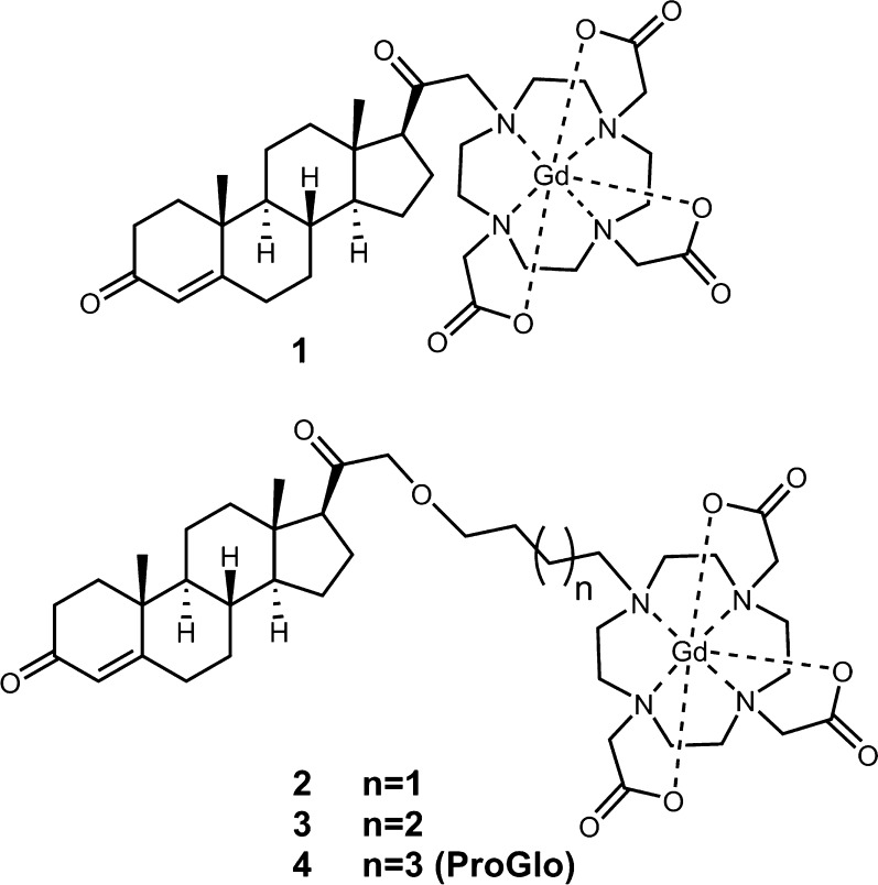
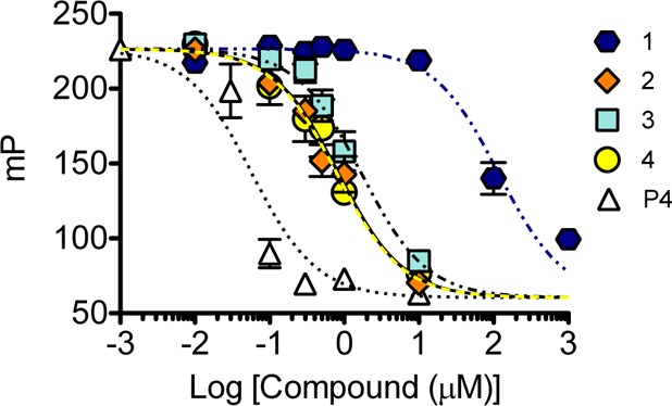
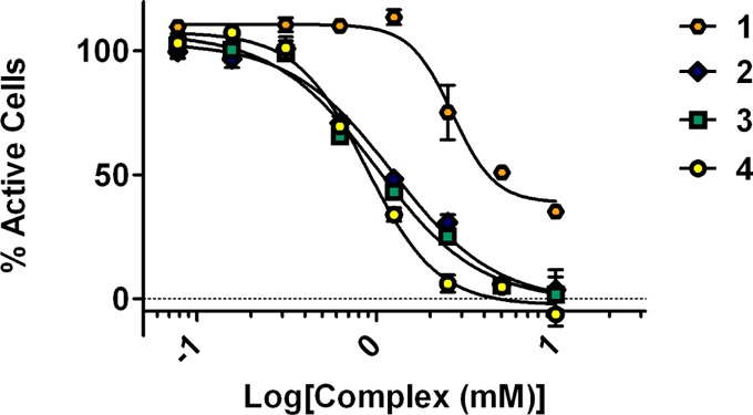
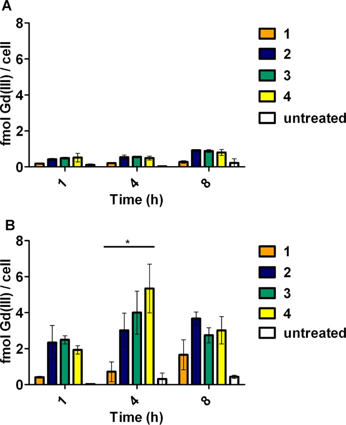
Determination of progesterone receptor (PR) status in hormone-dependent diseases is essential in ascertaining disease prognosis and monitoring treatment response. The development of a noninvasive means of monitoring these processes would have significant impact on early detection, cost, repeated measurements, and personalized treatment options. Magnetic resonance imaging (MRI) is widely recognized as a technique that can produce longitudinal studies, and PR-targeted MR probes may address a clinical problem by providing contrast enhancement that reports on PR status without biopsy. Commercially available MR contrast agents are typically delivered via intravenous injection, whereas steroids are administered subcutaneously. Whether the route of delivery is important for tissue accumulation of steroid-modified MRI contrast agents to PR-rich tissues is not known. To address this question, modification of the chemistry linking progesterone with the gadolinium chelate led to MR probes with increased water solubility and lower cellular toxicity and enabled administration through the blood. This attribute came at a cost through lower affinity for PR and decreased ability to cross the cell membrane, and ultimately it did not improve delivery of the PR-targeted MR probe to PR-rich tissues or tumors in vivo. Overall, these studies are important, as they demonstrate that targeted contrast agents require optimization of delivery and receptor binding of the steroid and the gadolinium chelate for optimal translation in vivo.
Figures
Similar articles
-
Water-Soluble Nanoconjugate for Enhanced Cellular Delivery of Receptor-Targeted Magnetic Resonance Contrast Agents.Bioconjug Chem. 2019 Nov 20;30(11):2947-2957. doi: 10.1021/acs.bioconjchem.9b00640. Epub 2019 Oct 22. Bioconjug Chem. 2019. PMID: 31589412 Free PMC article.
-
Synthesis and biological evaluation of water-soluble progesterone-conjugated probes for magnetic resonance imaging of hormone related cancers.Bioconjug Chem. 2011 Nov 16;22(11):2304-16. doi: 10.1021/bc2003555. Epub 2011 Oct 5. Bioconjug Chem. 2011. PMID: 21972997 Free PMC article.
-
A steroid-conjugated magnetic resonance probe enhances contrast in progesterone receptor expressing organs and tumors in vivo.Mol Pharm. 2011 Aug 1;8(4):1390-400. doi: 10.1021/mp200219e. Epub 2011 Jul 8. Mol Pharm. 2011. PMID: 21736390 Free PMC article.
-
Molecular Magnetic Resonance Imaging with Gd(III)-Based Contrast Agents: Challenges and Key Advances.J Am Chem Soc. 2019 Oct 30;141(43):17025-17041. doi: 10.1021/jacs.9b09149. Epub 2019 Oct 17. J Am Chem Soc. 2019. PMID: 31593630 Free PMC article. Review.
-
Progesterone receptor targeting with radiolabelled steroids: an approach in predicting breast cancer response to therapy.J Steroid Biochem Mol Biol. 2013 Sep;137:223-41. doi: 10.1016/j.jsbmb.2013.04.003. Epub 2013 May 10. J Steroid Biochem Mol Biol. 2013. PMID: 23669457 Review.
Cited by
-
Genome-wide transcriptional regulation of estrogen receptor targets in fallopian tube cells and the role of selective estrogen receptor modulators.J Ovarian Res. 2016 Feb 15;9:5. doi: 10.1186/s13048-016-0213-3. J Ovarian Res. 2016. PMID: 26879975 Free PMC article.
-
Water-Soluble Nanoconjugate for Enhanced Cellular Delivery of Receptor-Targeted Magnetic Resonance Contrast Agents.Bioconjug Chem. 2019 Nov 20;30(11):2947-2957. doi: 10.1021/acs.bioconjchem.9b00640. Epub 2019 Oct 22. Bioconjug Chem. 2019. PMID: 31589412 Free PMC article.
-
Estrogen Receptor-Targeted Contrast Agents for Molecular Magnetic Resonance Imaging of Breast Cancer Hormonal Status.Front Oncol. 2016 Apr 27;6:100. doi: 10.3389/fonc.2016.00100. eCollection 2016. Front Oncol. 2016. PMID: 27200289 Free PMC article.
References
-
- Czernin J.; Weber W. A.; Herschman H. R. (2006) Molecular imaging in the development of cancer therapeutics. Annu. Rev. Med. 57, 99–118. - PubMed
-
- Weissleder R. (2006) Molecular imaging in cancer. Science 312, 1168–1171. - PubMed
-
- Fukuda K.; Mori M.; Uchiyama M.; Iwai K.; Iwasaka T.; Sugimori H. (1998) Prognostic significance of progesterone receptor immunohistochemistry in endometrial carcinoma. Gynecol. Oncol. 69, 220–225. - PubMed
Publication types
MeSH terms
Substances
Grants and funding
LinkOut - more resources
Full Text Sources
Other Literature Sources
Medical
Research Materials
