. 2014 Jul 14;9(7):e100874.
doi: 10.1371/journal.pone.0100874. eCollection 2014.

Qualitative and quantitative comparison of the proteome of erythroid cells differentiated from human iPSCs and adult erythroid cells by multiplex TMT labelling and nanoLC-MS/MS

Affiliations

Qualitative and quantitative comparison of the proteome of erythroid cells differentiated from human iPSCs and adult erythroid cells by multiplex TMT labelling and nanoLC-MS/MS

Kongtana Trakarnsanga et al. PLoS One. .

Abstract

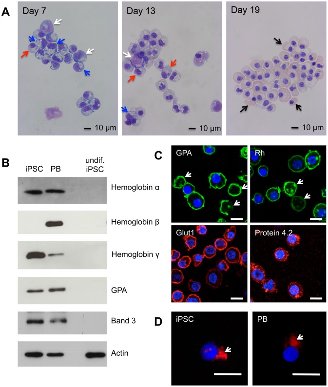
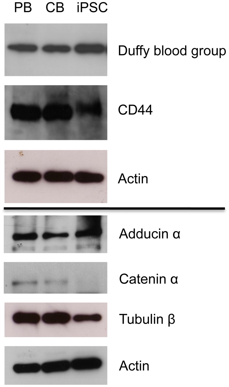
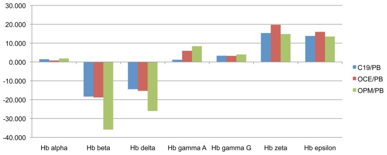
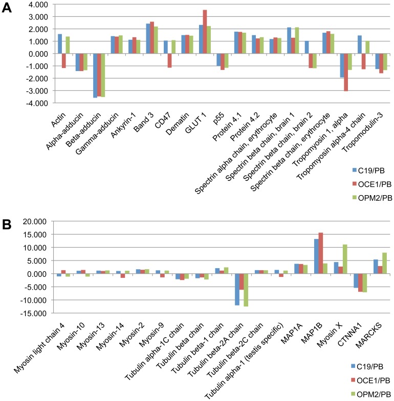
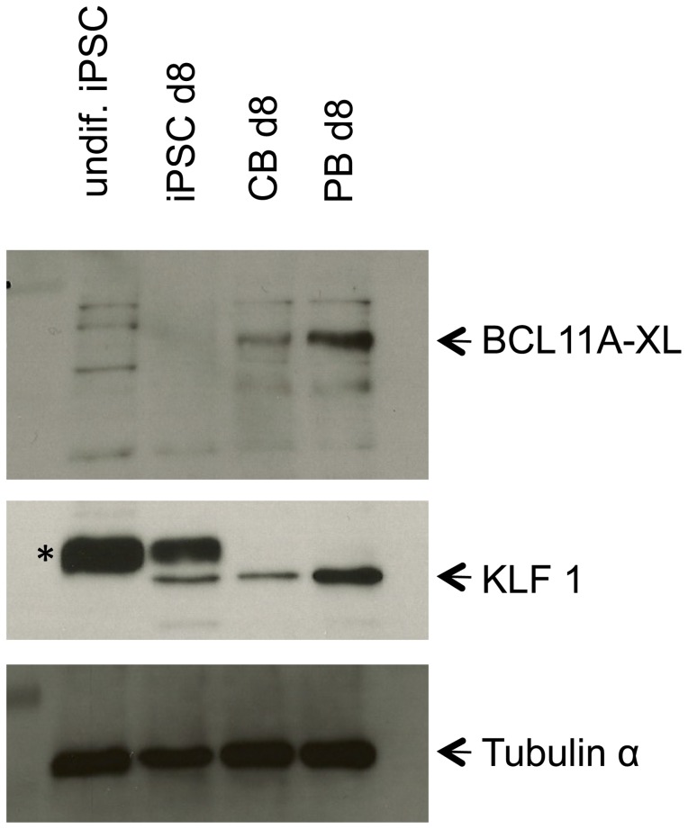
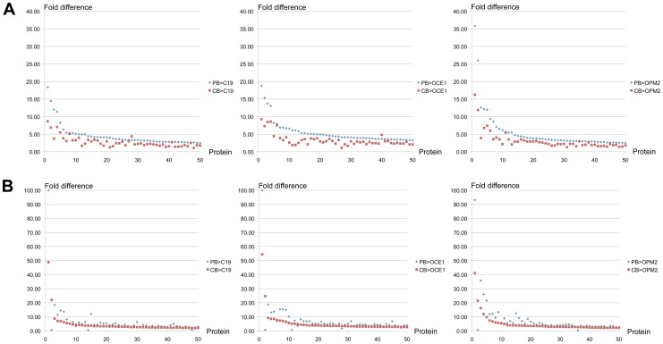
Induced pluripotent stem cells (iPSC) are an attractive progenitor source for the generation of in vitro blood products. However, before iPSC-derived erythroid cells can be considered for therapeutic use their similarity to adult erythroid cells must be confirmed. We have analysed the proteome of erythroid cells differentiated from the iPSC fibroblast derived line (C19) and showed they express hallmark RBC proteins, including all those of the ankyrin and 4.1R complex. We next compared the proteome of erythroid cells differentiated from three iPSC lines (C19, OCE1, OPM2) with that of adult and cord blood progenitors. Of the 1989 proteins quantified <3% differed in level by 2-fold or more between the different iPSC-derived erythroid cells. When compared to adult cells, 11% of proteins differed in level by 2-fold or more, falling to 1.9% if a 5-fold threshold was imposed to accommodate slight inter-cell line erythropoietic developmental variation. Notably, the level of >30 hallmark erythroid proteins was consistent between the iPSC lines and adult cells. In addition, a sub-population (10-15%) of iPSC erythroid cells in each of the iPSC lines completed enucleation. Aberrant expression of some cytoskeleton proteins may contribute to the failure of the majority of the cells to enucleate since we detected some alterations in cytoskeletal protein abundance. In conclusion, the proteome of erythroid cells differentiated from iPSC lines is very similar to that of normal adult erythroid cells, but further work to improve the induction of erythroid cells in existing iPSC lines or to generate novel erythroid cell lines is required before iPSC-derived red cells can be considered suitable for transfusion therapy.


Conflict of interest statement

Competing Interests: The authors have declared that no competing interests exist.

Figures

Figure 1
Figure 1. Erythroid differentiation of C19 iPSC CD34+ cells.
C19 and adult peripheral blood [PB] CD34+ cells were incubated for up to 21 days in our three-stage erythroid culture system. (A) Morphological analysis of cells stained with May-Grundwal Giemsa reagent on day 7, 13 and 19 in culture. Arrows, white proerythroblasts, blue basophillic erythroblasts, red polychromatic erythroblasts, black orthochromatic erythroblasts. (B) Western blot of iPSC and PB erythroid cells at day 19 in culture, and undifferentiated [undif] iPSCs, probed with antibodies to α-,β- and γ-globin, GPA and Band 3. Antibodies to actin were used as a protein loading control. Numbers on left are size markers. (C) iPSC erythroid cells at day 19 in culture probed with antibodies to GPA, Rh, GLUT1 and Protein 4.2, followed by compatible secondary antibodies with Alexa Fluor 488 (green) or Alexa Fluor 635 phalloidin (red). Arrows indicate reticulocytes. (D) Erythroid cells differentiated from C19 iPSC and PB progenitors were incubated with Alexa Fluor 635 phalloidin conjugated actin antibody (red). Arrows indicate contractile actin rings. Nuclear DNA was stained with blue-fluorescent DAPI. Images were obtained using a Leica SP5 confocal microscope. Scale bars 10 µm.
Figure 2
Figure 2. Levels of selected erythroid proteins in erythroid cells differentiated from iPSC, PB and CB.
Western blot of day 19 adult [PB], cord blood [CB] and C19 iPSC erythroid cell lysate (30 µg) probed with antibodies to Duffy and CD44, and day 8 lysate probed with antibodies to adducing α, catenin α (CTNNA1) and Tubulin β. Antibodies to actin were used as a protein loading control at both time points.
Figure 3
Figure 3. Difference in the level of globin subunits between erythroid cells differentiated in vitro from adult peripheral blood (PB) CD34+ cells, and from C19, OCE1 and OPM2 CD34+ cells.
PB, C19, OCE1 and OPM2 erythroid cells at day 8 in culture were lysed, proteins subjected to trypsin digest and resultant peptides labeled with isobaric tags for nanoLC-MS/MS based quantitation and comparison. Y-axis represents the fold difference in protein level between erythroid cells differentiated from each iPSC line and adult PB progenitors.
Figure 4
Figure 4. Difference in the level of RBC hallmark and cytoskeleton proteins between erythroid cells differentiated in vitro from adult peripheral blood (PB) CD34+ cells, and from C19, OCE1 and OPM2 CD34+ cells.
Erythroid cells differentiated from PB progenitors, C19, OCE1 and OPM2 iPSCs at day 8 in culture were lysed, proteins subjected to trypsin digest and resultant peptides labeled with isobaric tags for nanoLC-MS/MS based quantitation and comparison. Y-axis represents the fold difference in protein level between erythroid cells differentiated from each iPSC line and adult PB progenitors.
Figure 5
Figure 5. Expression of BCL11A and KLF1 in erythroid cells differentiated in vitro from C19 iPSC, cord blood (CB) and adult peripheral blood (PB) CD34+ cells.
Western blot of total protein from undifferentiated iPSCs (undif iPSC), and erythroid cells differentiated from iPSCs, CB and PB CD34+ cells at day 8 in culture probed with BCL11A antibody, stripped and re-probed with KLF1 antibody and stripped and re-probed with an antibody to tubulin as a protein loading control. * indicates non-specific band detected by antibody.
Figure 6
Figure 6. Difference in the level of proteins between adult (PB) and iPSC erythroid cells, compared to that between cord blood (CB) and iPSC erythroid cells.
PB, C19, OCE1 and OPM2 erythroid cells at day 8 in culture were lysed, proteins subjected to trypsin digest and resultant peptides labeled with isobaric tags for nanoLC-MS/MS based quantitation and comparison. The levels of 50 proteins with the greatest differential expression between (A) PB and C19, OCE1 or OPM2 erythroid cells and (B) CB and C19, OCE1 or OPM2 erythroid cells were all compared between PB and the 3 iPSC lines and CB and the 3 iPSC lines. Each point on the x-axis represents a unique protein. Y-axis represents the fold difference in protein level between erythroid cells differentiated from PB or CB and the iPSC line.

References

    1. Takahashi K, Yamanaka S (2006) Induction of pluripotent stem cells from mouse embryonic and adult fibroblast cultures by defined factors. Cell 126: 663–676. - PubMed
    1. Chang CJ, Mitra K, Koya M, Velho M, Desprat R, et al. (2011) Production of embryonic and fetal-like red blood cells from human induced pluripotent stem cells. PLoS One 6: e25761. - PMC - PubMed
    1. Hanna J, Wernig M, Markoulaki S, Sun CW, Meissner A, et al. (2007) Treatment of sickle cell anemia mouse model with iPS cells generated from autologous skin. Science 318: 1920–1923. - PubMed
    1. Seifinejad A, Taei A, Totonchi M, Vazirinasab H, Hassani SN, et al. (2010) Generation of human induced pluripotent stem cells from a Bombay individual: moving towards "universal-donor" red blood cells. Biochem Biophys Res Commun 391: 329–334. - PubMed
    1. Ye L, Chang JC, Lin C, Sun X, Yu J, et al. (2009) Induced pluripotent stem cells offer new approach to therapy in thalassemia and sickle cell anemia and option in prenatal diagnosis in genetic diseases. Proc Natl Acad Sci U S A 106: 9826–9830. - PMC - PubMed

Publication types

MeSH terms