. 2014 Jul-Aug;34(4):1082-98.
doi: 10.1148/rg.344140001.

Optimization of MR imaging for pretreatment evaluation of patients with endometrial and cervical cancer

Affiliations

Optimization of MR imaging for pretreatment evaluation of patients with endometrial and cervical cancer

Gaiane M Rauch et al. Radiographics. 2014 Jul-Aug.

Abstract

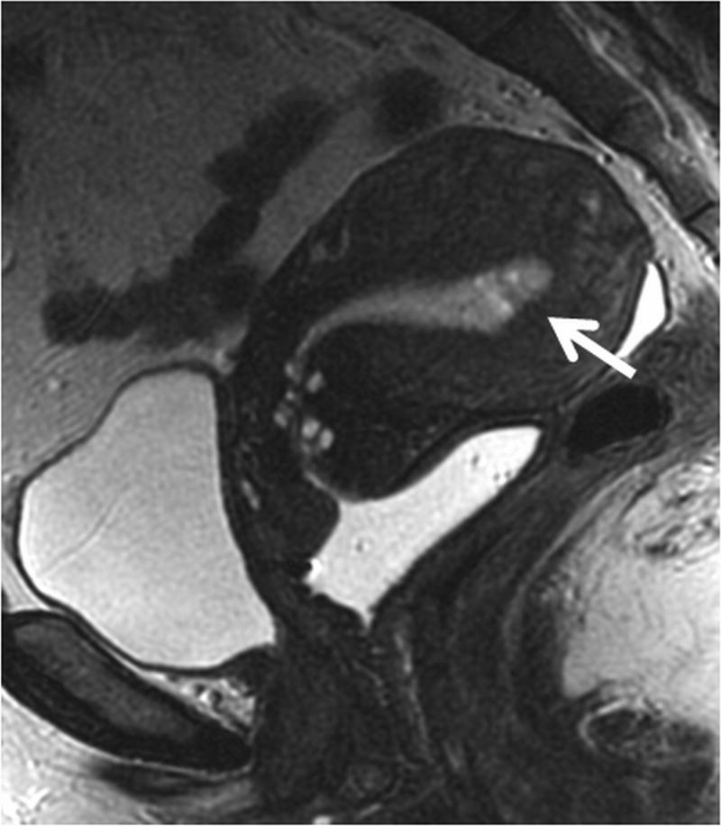
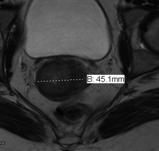
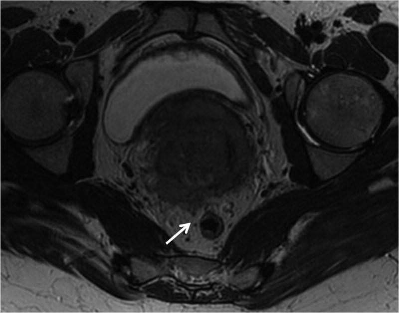
Endometrial and cervical cancer are the most common gynecologic malignancies in the world. Accurate staging of cervical and endometrial cancer is essential to determine the correct treatment approach. The current International Federation of Gynecology and Obstetrics (FIGO) staging system does not include modern imaging modalities. However, magnetic resonance (MR) imaging has proved to be the most accurate noninvasive modality for staging endometrial and cervical carcinomas and often helps with risk stratification and making treatment decisions. Multiparametric MR imaging is increasingly being used to evaluate the female pelvis, an approach that combines anatomic T2-weighted imaging with functional imaging (ie, dynamic contrast material-enhanced and diffusion-weighted imaging). MR imaging helps guide treatment decisions by depicting the depth of myometrial invasion and cervical stromal involvement in patients with endometrial cancer and tumor size and parametrial invasion in those with cervical cancer. However, its accuracy for local staging depends on technique and image quality, namely thin-section high-resolution multiplanar T2-weighted imaging with simple modifications, such as double oblique T2-weighting supplemented by diffusion weighting and contrast enhancement.


Figures

Figure 1
Figure 1. HR FRFSE T2-weighted images of the uterine zonal anatomy
Sagittal HR FRFSE T2 image shows the normal zonal anatomy of uterus. The high signal intensity endometrium is surrounded by the homogenous low signal intensity junctional zone (long arrow), which is continuous with the fibrous stroma of the cervix. The myometrium is of intermediate T2 signal (short arrow) and is contiguous with the outer interstitial stroma of the cervix.
Figure 2
Figure 2. Schematic of the “double oblique” sequence
The schematic shows uterus that is rotated anteriorly in the sagittal plane (anteverted uterus), but it is also tilted laterally to the left in the coronal plane. The double oblique sequence is obtained by angling images anteriorly in the sagittal plane (green line) and additionally angling images laterally in the coronal plane (blue line), which creates a “true oblique images” along the true axis of the uterus (orange line). A-anterior, P-posterior.
Figure 3
Figure 3. Double oblique MR imaging in endometrial cancer
(a) HR sagittal FRFSE T2-weighted image illustrates the correct plane for prescribing the orthogonal axial images perpendicular to the endometrial cavity in patients with endometrial carcinoma. The long line (small red arrow) marks the long axis of the uterus and dashed lines indicate the plane of acquisition of the routine oblique axial slices. (b) HR FRFSE Coronal T2-weighted image shows the body of the uterus is deviated to the right. The second oblique plane is prescribed perpendicular to the axis of the uterus in the coronal plane. The continuous line marks the axis of the endometrial cavity in the coronal plane and the dashed white lines illustrate the acquisition plane of the oblique axial images. The combination of both acquisitions prescribed along the long axis of the uterus in the sagittal and coronal planes form the double oblique axial image. (c) Routine oblique HR FRFSE T2-weighted image obtained as prescribed only on the sagittal images shows an apparent thinning of the right myometrium (arrow), which potentially could be mistaken for myometrial invasion. (d) Double oblique HR FRFSE T2-weighted image prescribed using both the sagittal and coronal planes is more appropriately angled along the true axis of the uterus, and shows the thickness of the myometrium to be symmetric (short arrows). Subtle superficial invasion of the inner myometrium is seen along the anterior wall (long arrow). The HR double oblique images are particularly useful when lateral deviation of the uterus is noted in the coronal plane, minimizing problems with volume averaging resulting from uterine position within the pelvis, which may lead to erroneous interpretations of myometrial invasion.
Figure 4
Figure 4. Suboptimal images from poor SNR can be improved by increasing FOV
(a) Coronal HR FRFSE T2-weighted image through the pelvis with 18 cm FOV do not clearly show the endometrial tumor due to poor SNR (arrow). (b) Coronal HR FRFSE T2-weighted images obtained with a larger FOV (increased from 18 to 20 cm). This simple step doubles the SNR and enables definition of the tumor seen expanding the endometrial cavity (arrow).
Figure 5
Figure 5. Optimizing image quality by eliminating artifacts and improving SNR, with anterior saturation band and adjusting the matrix size
(a) Oblique axial high resolution T2 weighted images through the uterus are degraded by motion artifact (arrow) and poor SNR, limiting evaluation of the endometrial-myometrial interface (matrix size 320×256). (b) Placement of an anterior saturation band (long arrow) to eliminate motion artifact from the anterior abdominal wall fat and a decrease in the matrix size (from 320×256 to 256×256) to increase the SNR improves image quality, permitting definition of a polypoid mass (short arrow), confined to the endometrial cavity.
Figure 6
Figure 6. Value of obtaining two orthogonal planes along the long axis of the tumor
(a) Sagittal HR FRFSE T2-weighted image shows a polypoid mass expanding the endometrial cavity (short arrow), extending into and expanding the endocervical canal (long arrow). It is difficult to state with certainty if there is myometrial invasion. (b) Axial HR FRFSE T2- weighted image along the coronal plane of the uterus, and orthogonal to the sagittal T2 images shows a broad based myometrial invasion along the right lateral wall (arrow), which is limited to the inner half of the myometrium. The depth of myometrial invasion was assessed using the thickness of the contralateral wall as a basis for assessment (double headed arrow). This was confirmed by pathology.
Figure 7
Figure 7. Adenomyosis can confound the assessment of myometrial invasion
(a) Sagittal HR FRFSE T2- weighted image shows widening of the junctional zone and small punctate foci of T2 hyperintensity and striations in the anterior myometrium reflecting adenomyosis (short arrow). The tumor is seen in the endometrial cavity (long arrow). (b) The sagittal 3D T1 post contrast images show defects in the anterior myometrium corresponding to the finding on the sagittal T2- weighted images (arrow). In this instance the signal intensity on the T2 weighted image and sharply marginated appearance on the post contrast image suggest adenomyosis in the anterior inner myometrium rather than tumor infiltration, but tumor infiltration cannot be excluded with certainty. The presence of adenomyosis limits the ability to accurately assess myometrial invasion on either sequence.
Figure 8
Figure 8. Value of Post Contrast T1 weighted images in Assessment of Myometrial Invasion
(a) Sagittal HR FRFSE T2-weighted image shows a small endometrial mass in the fundus of the uterus (arrow). (b) Sagittal dynamic 3D T1 FSPGR image shows myometrial invasion along the posterior uterine wall (arrow) confined to the inner myometrium. The delayed post contrast image (2-4 min) optimizes contrast between the myometrium and tumor.
Figure 9
Figure 9. Coregistration of HR FRFSE T2 and DCE-MRI utilizing same slice position
(a) High resolution axial T2 weighted images through the uterus show a hyperintense tumor in the endometrial cavity with a subtle area of signal abnormality seen in the anterior myometrium, suggesting possible invasion (short arrow). The images are degraded by motion artifact, which limits evaluation (long arrow). Note that an anterior saturation pulse was not placed over the anterior abdominal wall fat. (b) The delayed post contrast 3D T1 weighted images in the same plane confirm invasion into the inner myometrium (arrow). These images show the value of postcontrast images obtained in the same plane as the T2 weighted images in confirming a subtle finding, and providing a backup if one series is degraded by artifact and suboptimal for diagnostic evaluation.
Figure 10
Figure 10. Value of DW imaging obtained in comparable planes as the HR T2 FRFSE
(a) A HR FRFSE T2 weighted axial image that is coronal to the plane of the uterus because of angulation of uterus shows a tumor expanding the endometrial cavity with possible invasion of the right myometrium (arrow). (b) The oblique axial HR FRFSE T2 weighted images did not adequately delineate depth of invasion (arrow). (c and d) The DWI and ADC map image obtained with comparable obliquity shows restricted diffusion throughout the entire endometrium and confirms tumor infiltration into the right myometrium (short arrow). This case illustrates the value of obtaining DW imaging in an identical imaging plane to the FRFSE T2 images to increase confidence in assessment of depth of myometrial invasion. The endometrial cavity (long arrow) shows low ADC value compatible with tumor. Pathology confirmed tumor occupying the entire endometrial cavity and superficial invasion of the right myometrium.
Figure 11
Figure 11. Value of multiplanar HR MRI in adequate assessment of the tumor size
The measurements of the cervical tumor size should be obtained in all three orthogonal planes in order to achieve accurate size measurement. (a) Axial HR T2-weighted image size measurement of 4.5 cm underestimates tumor size in this patient with large cervical tumor. (b) Sagittal HR T2- weighted image demonstrates actual extent of the disease with largest measurement 6.1 cm.
Figure 12
Figure 12. HR MRI of locally advanced cervical cancer with bladder involvement (stage IVA)
(a) Sagittal HR FRFSE T2-weighted image shows a large mass replacing the entire cervix and invading the body of the uterus. Note extension of tumor into the bladder mucosa (short vertical arrow) and perirectal fat (long oblique arrow). (b) HR oblique FRFSE T2-weighted image shows irregular protrusions of tumor circumferentially infiltrating the parametria, consistent with bilateral parametrial extension, more prominent on the left (long horizontal arrow). Note invasion of the posterior wall of the urinary bladder with mucosal involvement on the left (short vertical arrow).
Figure 13
Figure 13. The value of orthogonal multiplanar HR FRFSE T2- weighted sequences for evaluation of cervical cancer
(a) High grade cervical adenocarcinoma seen as a hyperintense mass on the sagittal T2-weighted images (arrow). (b) The axial T2-weighted image is coronal to the plane of the tumor, limiting the evaluation for cervical stroma and parametrial invasion. The fibrous stroma on the right has an irregular contour (long arrow) and there are minute interruptions of the fibrous stroma on the left, suggesting deep cervical stromal invasion and possible parametrial extension (short arrows). (c) The oblique axial high resolution T2- weighted image perpendicular to the axis of the cervix, obtained at the level of possible deep cervical stromal invasion and parametrial extension, showed an intact thin layer of preserved fibrous stroma bilaterally (arrows), indicating the tumor is confined to the cervix.
Figure 14
Figure 14. Value of double-oblique HR FRFSE T2-weighted images for parametrial invasion evaluation in cervical cancer
(a) Sagittal HR FRFSE T2-weighted image shows an intermediate T2 signal intensity mass in the posterior cervical lip (arrow). The dashed line illustrates the acquisition plane of the oblique axial images obtained perpendicular to the long axis of the cervix, indicated by the continuous line. (b) Axial HR FRSE oblique T2-weighted image obtained based on the sagittal T2-weighted images shows the mass in the posterior cervical lip with suggestion of tumor infiltration into the left parametrium (arrow). (c) The cervix however is seen to be angled to the left of the mid line on the coronal T2 weighted images (arrow). The dashed line illustrates the angle of acquisition of the double oblique axial image perpendicular to the long axis of the laterally deviated cervix, which is indicated by the continuous line. (d) A double oblique T2- weighted image angled along the axis of the cervix based on the both the sagittal and coronal images shows an intact cervical stroma (arrow) between the tumor and the parametrium, excluding parametrial invasion on the left. This illustrates the value of double oblique images in eliminating the effects of volume averaging.
Figure 15
Figure 15. The value of 3DT2 sequence in cervical cancer imaging
(a) HR oblique FRFSE T2-weighted image shows a locally advanced cervical mass with bilateral parametrial extension (long arrows). The tumor invades the posterior cul-de-sac and abuts the anterior rectal wall (short arrow). (b) HR 3D T2-weighted image enables subsequent post-processing and image manipulation with alteration in the angle of the oblique axial image to show a clear fat plane between tumor and anterior rectal wall (arrow), which is an important factor for planning of the radiation therapy. The excellent T2 contrast of this sequence in addition to its multiplanar reformation capability makes it a valuable asset for evaluation of patients in equivocal cases. (c) The 3D T2-weighted images can be reviewed on any desirable imaging plane on a dedicated workstation. The multiplanar reformation capability of this sequence in addition to its excellent T2 contrast is very useful when additional anatomic detail is needed for the correct diagnosis and treatment planning.
Figure 16
Figure 16. HR MR Imaging with endovaginal gel
HR sagittal FRFSE T2-weighted sequence shows a large mass replacing the anterior and posterior cervical lips and protruding into the upper third of the vagina. The presence of high T2 signal endovaginal gel distends the vaginal fornices (arrows) and separates them from intravaginal exophytic component of the tumor. The vaginal wall and forniceal insertions are separate from the tumor, and there is preservation of the normal low T2 signal intensity of the muscular vaginal wall, excluding vaginal invasion.
Figure 17
Figure 17. HR MR imaging of metastatic lymphadenopathy
Coronal HR FRFSE T2-weighted image shows a large cervical mass with bilateral parametrial extension (short horizontal arrows). Bilateral external iliac lymphadenopathy is identified (long vertical arrows). The nodes display an abnormal signal intensity that matches the signal intensity of the primary cervical tumor. HR MR imaging is useful to demonstrate abnormal alterations in nodal morphology and signal intensity, even in normal sized lymph nodes, increasing the sensitivity for nodal disease detection.

References

    1. Howlader N, Noone A, Krapcho M, et al. SEER Cancer Statistics Review (CSR), 1975–2010. [Accessed April 8, 2013];National Cancer Institute [serial online] 2012 Available at: http://seer.cancer.gov/csr/1975_2010/
    1. Sala E, Wakely S, Senior E, Lomas D. MRI of malignant neoplasms of the uterine corpus and cervix. AJR Am J Roentgenol. 2007;188(6):1577–1587. - PubMed
    1. Lewin SN. Revised FIGO staging system for endometrial cancer. Clin Obstet Gynecol. 2011;54(2):215–218. - PubMed
    1. Sala E, Rockall A, Kubik-Huch RA. Advances in magnetic resonance imaging of endometrial cancer. Eur Radiol. 2011;21(3):468–473. - PubMed
    1. Larson DM, Connor GP, Broste SK, Krawisz BR, Johnson KK. Prognostic significance of gross myometrial invasion with endometrial cancer. Obstet Gynecol. 1996;88(3):394–398. - PubMed

MeSH terms