. 2014 Jul-Aug;34(4):E89-102.
doi: 10.1148/rg.344130087.

Digital breast tomosynthesis: lessons learned from early clinical implementation

Affiliations

Digital breast tomosynthesis: lessons learned from early clinical implementation

Robyn Gartner Roth et al. Radiographics. 2014 Jul-Aug.

Abstract

The limitations of mammography are well known and are partly related to the fact that with conventional imaging, the three-dimensional volume of the breast is imaged and presented in a two-dimensional format. Because normal breast tissue is similar in x-ray attenuation to some breast cancers, clinically relevant malignancies may be obscured by normal overlapping tissue. In addition, complex areas of normal tissue may be perceived as suspicious. The limitations of two-dimensional breast imaging lead to low sensitivity in detecting some cancers and high false-positive recall rates. Although mammographic screening has been shown to reduce breast cancer deaths by approximately 30%, controversy exists over when and how often screening mammography should occur. Digital breast tomosynthesis (DBT) is rapidly being implemented in breast imaging clinics around the world as early clinical data demonstrate that it may address some of the limitations of conventional mammography. With DBT, multiple low-dose x-ray images are acquired in an arc and reconstructed to create a three-dimensional image, thus minimizing the impact of overlapping breast tissue and improving lesion conspicuity. Early studies of screening DBT have shown decreased false-positive callback rates and increased rates of cancer detection (particularly for invasive cancers), resulting in increased sensitivity and specificity. In our clinical practice, we have completed more than 2 years of using two-view digital mammography combined with two-view DBT for all screening and select diagnostic imaging examinations (over 25,000 patients). Our experience, combined with previously published data, demonstrates that the combined use of DBT and digital mammography is associated with improved outcomes for screening and diagnostic imaging. Online supplemental material is available for this article.


Figures

Figure 1
Figure 1
Drawing shows how DBT images are obtained. Multiple low-dose x-ray projection images are acquired in an arc and used to reconstruct a 3D image of the breast.
Figure 2a
Figure 2a
Invasive lobular carcinoma in a 68-year-old woman. (a, b) Findings on bilateral MLO (a) and craniocaudal (b) 2D screening mammograms were initially interpreted as negative. Corresponding DBT images (Movies 1, 2) show architectural distortion and an irregular mass in the upper outer right breast, findings not seen at DM. (c) US image of the right breast shows an irregular mass at the 10-o’clock position, 5 cm from the nipple. (d) Sagittal contrast-enhanced fat-saturated T1-weighted magnetic resonance (MR) subtraction image of the right breast helps confirm the DBT findings. Pathologic analysis demonstrated invasive lobular carcinoma.
Figure 2b
Figure 2b
Invasive lobular carcinoma in a 68-year-old woman. (a, b) Findings on bilateral MLO (a) and craniocaudal (b) 2D screening mammograms were initially interpreted as negative. Corresponding DBT images (Movies 1, 2) show architectural distortion and an irregular mass in the upper outer right breast, findings not seen at DM. (c) US image of the right breast shows an irregular mass at the 10-o’clock position, 5 cm from the nipple. (d) Sagittal contrast-enhanced fat-saturated T1-weighted magnetic resonance (MR) subtraction image of the right breast helps confirm the DBT findings. Pathologic analysis demonstrated invasive lobular carcinoma.
Figure 2c
Figure 2c
Invasive lobular carcinoma in a 68-year-old woman. (a, b) Findings on bilateral MLO (a) and craniocaudal (b) 2D screening mammograms were initially interpreted as negative. Corresponding DBT images (Movies 1, 2) show architectural distortion and an irregular mass in the upper outer right breast, findings not seen at DM. (c) US image of the right breast shows an irregular mass at the 10-o’clock position, 5 cm from the nipple. (d) Sagittal contrast-enhanced fat-saturated T1-weighted magnetic resonance (MR) subtraction image of the right breast helps confirm the DBT findings. Pathologic analysis demonstrated invasive lobular carcinoma.
Figure 2d
Figure 2d
Invasive lobular carcinoma in a 68-year-old woman. (a, b) Findings on bilateral MLO (a) and craniocaudal (b) 2D screening mammograms were initially interpreted as negative. Corresponding DBT images (Movies 1, 2) show architectural distortion and an irregular mass in the upper outer right breast, findings not seen at DM. (c) US image of the right breast shows an irregular mass at the 10-o’clock position, 5 cm from the nipple. (d) Sagittal contrast-enhanced fat-saturated T1-weighted magnetic resonance (MR) subtraction image of the right breast helps confirm the DBT findings. Pathologic analysis demonstrated invasive lobular carcinoma.
Figure 3a
Figure 3a
Invasive ductal carcinoma (IDC) in a 72-year-old woman. (a) Findings on these bilateral craniocaudal screening DM images were initially interpreted as negative, but DBT images (Movie 3) show a spiculated mass in the medial right breast. (b) US image of the right medial breast shows an irregular hypoechoic mass with shadowing, a finding that corresponds to the abnormality seen at DBT. Pathologic analysis revealed a 5-mm IDC.
Figure 3b
Figure 3b
Invasive ductal carcinoma (IDC) in a 72-year-old woman. (a) Findings on these bilateral craniocaudal screening DM images were initially interpreted as negative, but DBT images (Movie 3) show a spiculated mass in the medial right breast. (b) US image of the right medial breast shows an irregular hypoechoic mass with shadowing, a finding that corresponds to the abnormality seen at DBT. Pathologic analysis revealed a 5-mm IDC.
Figure 4a
Figure 4a
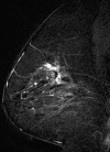
IDC in a 42-year-old woman. (a, b) Bilateral MLO (a) and craniocaudal (b) screening DM images show subtle architectural distortion in the lateral and subareolar right breast, a finding that is more conspicuous on DBT images (Movies 4, 5). The extensive nature of the finding is also appreciated on the DBT images. (c) Sagittal contrast-enhanced fat-saturated T1-weighted MR subtraction image of the right breast shows patchy extensive enhancement in the region of architectural distortion. Pathologic analysis confirmed extensive IDC.
Figure 4b
Figure 4b
IDC in a 42-year-old woman. (a, b) Bilateral MLO (a) and craniocaudal (b) screening DM images show subtle architectural distortion in the lateral and subareolar right breast, a finding that is more conspicuous on DBT images (Movies 4, 5). The extensive nature of the finding is also appreciated on the DBT images. (c) Sagittal contrast-enhanced fat-saturated T1-weighted MR subtraction image of the right breast shows patchy extensive enhancement in the region of architectural distortion. Pathologic analysis confirmed extensive IDC.
Figure 4c
Figure 4c
IDC in a 42-year-old woman. (a, b) Bilateral MLO (a) and craniocaudal (b) screening DM images show subtle architectural distortion in the lateral and subareolar right breast, a finding that is more conspicuous on DBT images (Movies 4, 5). The extensive nature of the finding is also appreciated on the DBT images. (c) Sagittal contrast-enhanced fat-saturated T1-weighted MR subtraction image of the right breast shows patchy extensive enhancement in the region of architectural distortion. Pathologic analysis confirmed extensive IDC.
Figure 5
Figure 5
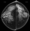
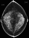
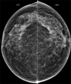
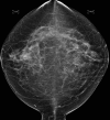
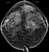
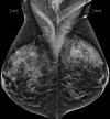
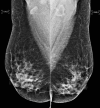
Craniocaudal screening DM images in a 55-year-old woman show a focal asymmetry in the lateral right breast. DBT images (Movie 6) demonstrate the abnormality to be related to overlapping breast tissue. No further workup is needed.
Figure 6
Figure 6
Craniocaudal screening DM images in a 47-year-old woman show a focal asymmetry in the lateral left breast; corresponding DBT images (Movie 7) show no abnormality. The findings at DM are related to tissue superimposition. No further workup is needed.
Figure 7
Figure 7
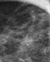
Findings on these MLO screening DM images of the right breast in a 62-year-old woman were initially interpreted as negative; however, a subtle architectural distortion was identified in the upper right breast on DBT images (Movie 8). Pathologic analysis revealed a radial scar with atypical ductal hyperplasia.
Figure 8a
Figure 8a
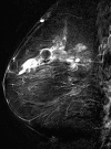
DCIS and invasive lobular cancer in a 45-year-old woman. (a–c) Suspicious calcifications are seen in the upper outer right breast on craniocaudal (a) and MLO (b) screening DM images. The calcifications are also seen on a zoomed-in DM image (c). DBT images show calcifications in the right breast (Movies 9, 10) and also depict a cyst in the left breast, which otherwise is normal (Movie 11). Pathologic analysis revealed a 2-cm DCIS in the right breast. (d, e) Sagittal contrast-enhanced fat-saturated T1-weighted MR subtraction images of the right (d) and left (e) breasts obtained for staging show enhancement of the known DCIS in the right breast and extensive suspicious enhancement in the left breast, a finding that was originally interpreted as negative on the DM and DBT images. Pathologic analysis showed a 6-cm invasive lobular cancer in the left breast, with extensive lobular carcinoma in situ.
Figure 8b
Figure 8b
DCIS and invasive lobular cancer in a 45-year-old woman. (a–c) Suspicious calcifications are seen in the upper outer right breast on craniocaudal (a) and MLO (b) screening DM images. The calcifications are also seen on a zoomed-in DM image (c). DBT images show calcifications in the right breast (Movies 9, 10) and also depict a cyst in the left breast, which otherwise is normal (Movie 11). Pathologic analysis revealed a 2-cm DCIS in the right breast. (d, e) Sagittal contrast-enhanced fat-saturated T1-weighted MR subtraction images of the right (d) and left (e) breasts obtained for staging show enhancement of the known DCIS in the right breast and extensive suspicious enhancement in the left breast, a finding that was originally interpreted as negative on the DM and DBT images. Pathologic analysis showed a 6-cm invasive lobular cancer in the left breast, with extensive lobular carcinoma in situ.
Figure 8c
Figure 8c
DCIS and invasive lobular cancer in a 45-year-old woman. (a–c) Suspicious calcifications are seen in the upper outer right breast on craniocaudal (a) and MLO (b) screening DM images. The calcifications are also seen on a zoomed-in DM image (c). DBT images show calcifications in the right breast (Movies 9, 10) and also depict a cyst in the left breast, which otherwise is normal (Movie 11). Pathologic analysis revealed a 2-cm DCIS in the right breast. (d, e) Sagittal contrast-enhanced fat-saturated T1-weighted MR subtraction images of the right (d) and left (e) breasts obtained for staging show enhancement of the known DCIS in the right breast and extensive suspicious enhancement in the left breast, a finding that was originally interpreted as negative on the DM and DBT images. Pathologic analysis showed a 6-cm invasive lobular cancer in the left breast, with extensive lobular carcinoma in situ.
Figure 8d
Figure 8d
DCIS and invasive lobular cancer in a 45-year-old woman. (a–c) Suspicious calcifications are seen in the upper outer right breast on craniocaudal (a) and MLO (b) screening DM images. The calcifications are also seen on a zoomed-in DM image (c). DBT images show calcifications in the right breast (Movies 9, 10) and also depict a cyst in the left breast, which otherwise is normal (Movie 11). Pathologic analysis revealed a 2-cm DCIS in the right breast. (d, e) Sagittal contrast-enhanced fat-saturated T1-weighted MR subtraction images of the right (d) and left (e) breasts obtained for staging show enhancement of the known DCIS in the right breast and extensive suspicious enhancement in the left breast, a finding that was originally interpreted as negative on the DM and DBT images. Pathologic analysis showed a 6-cm invasive lobular cancer in the left breast, with extensive lobular carcinoma in situ.
Figure 8e
Figure 8e
DCIS and invasive lobular cancer in a 45-year-old woman. (a–c) Suspicious calcifications are seen in the upper outer right breast on craniocaudal (a) and MLO (b) screening DM images. The calcifications are also seen on a zoomed-in DM image (c). DBT images show calcifications in the right breast (Movies 9, 10) and also depict a cyst in the left breast, which otherwise is normal (Movie 11). Pathologic analysis revealed a 2-cm DCIS in the right breast. (d, e) Sagittal contrast-enhanced fat-saturated T1-weighted MR subtraction images of the right (d) and left (e) breasts obtained for staging show enhancement of the known DCIS in the right breast and extensive suspicious enhancement in the left breast, a finding that was originally interpreted as negative on the DM and DBT images. Pathologic analysis showed a 6-cm invasive lobular cancer in the left breast, with extensive lobular carcinoma in situ.
Figure 9a
Figure 9a
IDC with DCIS in a 57-year-old woman. (a, b) Craniocaudal (a) and MLO (b) screening DM images show a questionable focal asymmetry in the upper outer left breast. DBT images of the left breast (Movies 12, 13) show no abnormality, and no abnormality was seen on corresponding US images (not shown). The finding on the DM images is due to superimposition. However, a suspicious area is seen in the upper posterior right breast on DBT images (Movie 14). (c) Spot compression image of the right breast shows no definite abnormality because of tissue effacement. (d) US image of the right breast shows a hypoechoic mass at the 12-o’clock position, 2 cm from the areolar margin, a finding that corresponds to the abnormality seen at DBT. Pathologic analysis revealed an 8-mm IDC with DCIS in the right breast.
Figure 9b
Figure 9b
IDC with DCIS in a 57-year-old woman. (a, b) Craniocaudal (a) and MLO (b) screening DM images show a questionable focal asymmetry in the upper outer left breast. DBT images of the left breast (Movies 12, 13) show no abnormality, and no abnormality was seen on corresponding US images (not shown). The finding on the DM images is due to superimposition. However, a suspicious area is seen in the upper posterior right breast on DBT images (Movie 14). (c) Spot compression image of the right breast shows no definite abnormality because of tissue effacement. (d) US image of the right breast shows a hypoechoic mass at the 12-o’clock position, 2 cm from the areolar margin, a finding that corresponds to the abnormality seen at DBT. Pathologic analysis revealed an 8-mm IDC with DCIS in the right breast.
Figure 9c
Figure 9c
IDC with DCIS in a 57-year-old woman. (a, b) Craniocaudal (a) and MLO (b) screening DM images show a questionable focal asymmetry in the upper outer left breast. DBT images of the left breast (Movies 12, 13) show no abnormality, and no abnormality was seen on corresponding US images (not shown). The finding on the DM images is due to superimposition. However, a suspicious area is seen in the upper posterior right breast on DBT images (Movie 14). (c) Spot compression image of the right breast shows no definite abnormality because of tissue effacement. (d) US image of the right breast shows a hypoechoic mass at the 12-o’clock position, 2 cm from the areolar margin, a finding that corresponds to the abnormality seen at DBT. Pathologic analysis revealed an 8-mm IDC with DCIS in the right breast.
Figure 9d
Figure 9d
IDC with DCIS in a 57-year-old woman. (a, b) Craniocaudal (a) and MLO (b) screening DM images show a questionable focal asymmetry in the upper outer left breast. DBT images of the left breast (Movies 12, 13) show no abnormality, and no abnormality was seen on corresponding US images (not shown). The finding on the DM images is due to superimposition. However, a suspicious area is seen in the upper posterior right breast on DBT images (Movie 14). (c) Spot compression image of the right breast shows no definite abnormality because of tissue effacement. (d) US image of the right breast shows a hypoechoic mass at the 12-o’clock position, 2 cm from the areolar margin, a finding that corresponds to the abnormality seen at DBT. Pathologic analysis revealed an 8-mm IDC with DCIS in the right breast.
Figure 10a
Figure 10a
IDC in a 77-year-old woman. (a, b) Findings on bilateral craniocaudal (a) and MLO (b) screening DM images were initially interpreted as normal. On craniocaudal DBT images, a subtle area of architectural distortion is seen in the lateral right breast, midway through the stack (Movie 15), and is triangulated to the mid breast on MLO and mediolateral DBT images (Movies 16, 17). (c, d) On spot compression images of the right breast, the lesion is effaced and not readily visible. (e) Targeted US image of the right breast obtained on the basis of the triangulated location from the DBT images depicts the lesion. Pathologic analysis revealed IDC.
Figure 10b
Figure 10b
IDC in a 77-year-old woman. (a, b) Findings on bilateral craniocaudal (a) and MLO (b) screening DM images were initially interpreted as normal. On craniocaudal DBT images, a subtle area of architectural distortion is seen in the lateral right breast, midway through the stack (Movie 15), and is triangulated to the mid breast on MLO and mediolateral DBT images (Movies 16, 17). (c, d) On spot compression images of the right breast, the lesion is effaced and not readily visible. (e) Targeted US image of the right breast obtained on the basis of the triangulated location from the DBT images depicts the lesion. Pathologic analysis revealed IDC.
Figure 10c
Figure 10c
IDC in a 77-year-old woman. (a, b) Findings on bilateral craniocaudal (a) and MLO (b) screening DM images were initially interpreted as normal. On craniocaudal DBT images, a subtle area of architectural distortion is seen in the lateral right breast, midway through the stack (Movie 15), and is triangulated to the mid breast on MLO and mediolateral DBT images (Movies 16, 17). (c, d) On spot compression images of the right breast, the lesion is effaced and not readily visible. (e) Targeted US image of the right breast obtained on the basis of the triangulated location from the DBT images depicts the lesion. Pathologic analysis revealed IDC.
Figure 10d
Figure 10d
IDC in a 77-year-old woman. (a, b) Findings on bilateral craniocaudal (a) and MLO (b) screening DM images were initially interpreted as normal. On craniocaudal DBT images, a subtle area of architectural distortion is seen in the lateral right breast, midway through the stack (Movie 15), and is triangulated to the mid breast on MLO and mediolateral DBT images (Movies 16, 17). (c, d) On spot compression images of the right breast, the lesion is effaced and not readily visible. (e) Targeted US image of the right breast obtained on the basis of the triangulated location from the DBT images depicts the lesion. Pathologic analysis revealed IDC.
Figure 10e
Figure 10e
IDC in a 77-year-old woman. (a, b) Findings on bilateral craniocaudal (a) and MLO (b) screening DM images were initially interpreted as normal. On craniocaudal DBT images, a subtle area of architectural distortion is seen in the lateral right breast, midway through the stack (Movie 15), and is triangulated to the mid breast on MLO and mediolateral DBT images (Movies 16, 17). (c, d) On spot compression images of the right breast, the lesion is effaced and not readily visible. (e) Targeted US image of the right breast obtained on the basis of the triangulated location from the DBT images depicts the lesion. Pathologic analysis revealed IDC.
Figure 11a
Figure 11a
IDC in a 60-year-old woman. (a, b) Findings on bilateral craniocaudal (a) and MLO (b) screening DM images were initially interpreted as negative. DBT images show a subtle area of architectural distortion in the mid left breast, a finding best seen on an MLO DBT image (Movie 18). The distortion is not definitely seen on craniocaudal DBT images (Movie 19). Since the MLO DBT stack goes from medial to lateral and the lesion is viewed toward the end of the stack, the lesion was triangulated from the MLO view to the lateral mid breast. The lesion then could be localized on craniocaudal DBT images. Spot magnification images showed no abnormality, but the ability to triangulate from the DBT stack guided US evaluation. (c) US image of the left breast shows an irregular hypoechoic mass at the 3-o’clock position, 5 cm from the areolar margin, a finding that corresponds to the architectural distortion seen at DBT. Pathologic analysis revealed a 4-mm intermediate-grade IDC.
Figure 11b
Figure 11b
IDC in a 60-year-old woman. (a, b) Findings on bilateral craniocaudal (a) and MLO (b) screening DM images were initially interpreted as negative. DBT images show a subtle area of architectural distortion in the mid left breast, a finding best seen on an MLO DBT image (Movie 18). The distortion is not definitely seen on craniocaudal DBT images (Movie 19). Since the MLO DBT stack goes from medial to lateral and the lesion is viewed toward the end of the stack, the lesion was triangulated from the MLO view to the lateral mid breast. The lesion then could be localized on craniocaudal DBT images. Spot magnification images showed no abnormality, but the ability to triangulate from the DBT stack guided US evaluation. (c) US image of the left breast shows an irregular hypoechoic mass at the 3-o’clock position, 5 cm from the areolar margin, a finding that corresponds to the architectural distortion seen at DBT. Pathologic analysis revealed a 4-mm intermediate-grade IDC.
Figure 11c
Figure 11c
IDC in a 60-year-old woman. (a, b) Findings on bilateral craniocaudal (a) and MLO (b) screening DM images were initially interpreted as negative. DBT images show a subtle area of architectural distortion in the mid left breast, a finding best seen on an MLO DBT image (Movie 18). The distortion is not definitely seen on craniocaudal DBT images (Movie 19). Since the MLO DBT stack goes from medial to lateral and the lesion is viewed toward the end of the stack, the lesion was triangulated from the MLO view to the lateral mid breast. The lesion then could be localized on craniocaudal DBT images. Spot magnification images showed no abnormality, but the ability to triangulate from the DBT stack guided US evaluation. (c) US image of the left breast shows an irregular hypoechoic mass at the 3-o’clock position, 5 cm from the areolar margin, a finding that corresponds to the architectural distortion seen at DBT. Pathologic analysis revealed a 4-mm intermediate-grade IDC.
Figure 12
Figure 12
DBT used to localize calcifications to the skin. Craniocaudal DM image of the left breast shows clustered calcifications, findings that are clearly visible in the skin layer on the DBT images (Movie 20).

Comment in

References

    1. Tabár L, Vitak B, Chen TH, et al. . Swedish Two-County Trial: impact of mammographic screening on breast cancer mortality during 3 decades. Radiology 2011;260(3):658–663. - PubMed
    1. Pisano ED, Gatsonis C, Hendrick E, et al. . Diagnostic performance of digital versus film mammography for breast-cancer screening. N Engl J Med 2005;353(17):1773–1783. - PubMed
    1. U.S. Preventive Services Task Force . Screening for breast cancer: U.S. Preventive Services Task Force recommendation statement. Ann Intern Med 2009; 151(10):716–726, W236. - PubMed
    1. ACR Practice Guideline for the Performance of Screening and Diagnostic Mammography. American College of Radiology Web site. http://www.acr.org/∼/media/ACR/Documents/PGTS/guidelines/Screening_Mammo.... Updated 2008. Accessed May 20, 2013.
    1. American College of Obstetricians-Gynecologists . Practice Bulletin No. 122: Breast cancer screening. Obstet Gynecol 2011;118(2 Pt 1):372–382. - PubMed

LinkOut - more resources