. 2014 Sep 26:277:250-66.
doi: 10.1016/j.neuroscience.2014.07.006. Epub 2014 Jul 11.

Bidirectional modulation of deep cerebellar nuclear cells revealed by optogenetic manipulation of inhibitory inputs from Purkinje cells

Affiliations

Bidirectional modulation of deep cerebellar nuclear cells revealed by optogenetic manipulation of inhibitory inputs from Purkinje cells

V Z Han et al. Neuroscience. .

Abstract

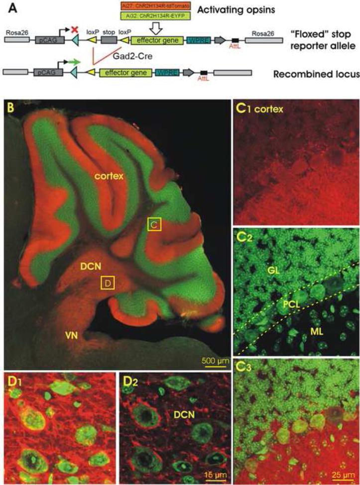
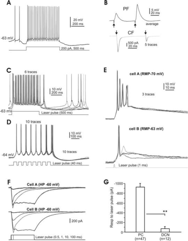
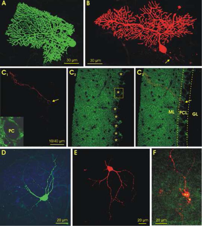
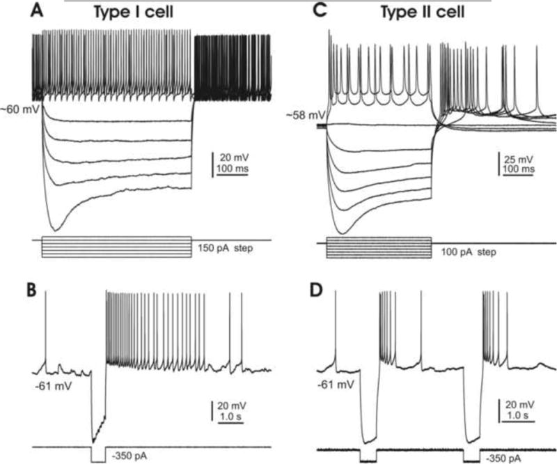
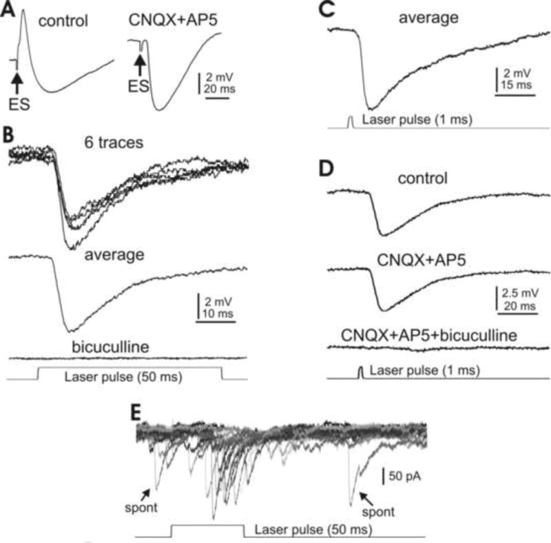
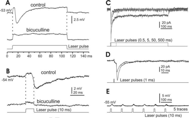
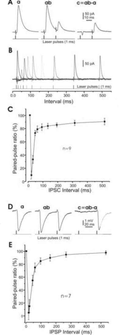
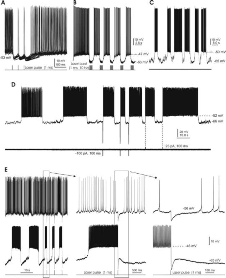
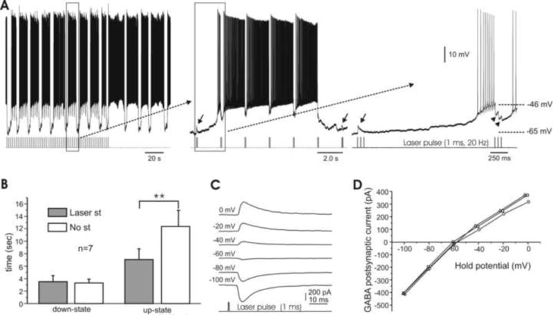
In the mammalian cerebellum, deep cerebellar nuclear (DCN) cells convey all information from cortical Purkinje cells (PCs) to premotor nuclei and other brain regions. However, how DCN cells integrate inhibitory input from PCs with excitatory inputs from other sources has been difficult to assess, in part due to the large spatial separation between cortical PCs and their target cells in the nuclei. To circumvent this problem we have used a Cre-mediated genetic approach to generate mice in which channelrhodopsin-2 (ChR2), fused with a fluorescent reporter, is selectively expressed by GABAergic neurons, including PCs. In recordings from brain slice preparations from this model, mammalian PCs can be robustly depolarized and discharged by brief photostimulation. In recordings of postsynaptic DCN cells, photostimulation of PC axons induces a strong inhibition that resembles these cells' responses to focal electrical stimulation, but without a requirement for the glutamate receptor blockers typically applied in such experiments. In this optogenetic model, laser pulses as brief as 1 ms can reliably induce an inhibition that shuts down the spontaneous spiking of a DCN cell for ∼50 ms. If bursts of such brief light pulses are delivered, a fixed pattern of bistable bursting emerges. If these pulses are delivered continuously to a spontaneously bistable cell, the immediate response to such photostimulation is inhibitory in the cell's depolarized state and excitatory when the membrane has repolarized; a less regular burst pattern then persists after stimulation has been terminated. These results indicate that the spiking activity of DCN cells can be bidirectionally modulated by the optically activated synaptic inhibition of cortical PCs.

Keywords: Purkinje cell; deep nuclear cell; optogenetic model; photostimulation; synaptic inhibition.


Figures

Fig. 1
Fig. 1. Generation and characterization of Gad2Cre/Ai27 and Gad2Cre/Ai32 mice expressing tdTomato- or EYFP-ChR2 in GABAergic cerebellar neurons
A. Schematic for the activation of an inducible ChR2-tdTomato or ChR2-EYFP gene targeted to the Rosa26 locus using Gad2Cre. B. Image of the Gad2Cre/Ai27 mouse cerebellum in a parasagittal plane, showing tdTomato (red) counterstained with Nissl fluorescence (green). DCN, deep cerebellar nuclei; VN, vestibular nuclei. C. Confocal images of boxed area C of the cerebellar cortex in B, showing strong ChR2-tdTomato fluorescence in both somas and dendrites of PCs (C1), Nissl staining with green fluorescent dye (C2), and their overlay (C3). GL, granular layer; ML, molecular layer; PCL, Purkinje cell layer. D. Confocal image of a DCN region in B (boxed area D) in a Z-stack (10.5 μm, D1) and in a single optical section (0.5 μm, D2), showing that cells are heavily innervated by ChR2-positive fibers. Scale bar: 500 μm in B, 25 μm in C, and 15 μm in D.
Fig. 2
Fig. 2. Physiology of Purkinje cells in Gad2Cre/Ai27(32) mice
A. Typical spiking of a Purkinje cell, spontaneously and in response to an inward current step. B. Responses of the same cell shown in A to parallel fiber (PF, top) and climbing fiber (CF, bottom) activation. Note the respective paired-pulse facilitation and depression of the PF and CF responses. C. Evoked burst spiking at ∼25 Hz elicited by a 500 ms laser pulse, followed by spontaneous spiking. D. Shorter (40 ms) laser pulses evoke spiking at 25 Hz. E. Dual recordings of a pair of PCs under current-clamp conditions. A single brief (1 ms) laser pulse reliably elicits a large depolarization and spikes in both cells. Note the complex spike-like responses in cell A. F. Dual recordings of a pair of PCs in voltage-clamp mode, showing that laser pulses at variable durations evoke robust inward currents consisting of phasic and plateau components in both cells. G. Plateau current responses of different cell types to longer laser pulses (≥50 ms). RMP, resting membrane potential; PC, Purkinje cells; DCN, (small- and medium-sized) deep cerebellar nuclear cells. **, P<0.01. Note that here and in the following figures, single or average traces are shown unless otherwise specified.
Fig. 3
Fig. 3. Morphology of cerebellar cortical and deep nuclear cells filled with intracellular tracers following their physiological characterization
A&B. Confocal images of two Purkinje cells in parasagittal slices, labeled with green fluorescent dye in a Gad2Cre/Ai27 mouse (with a tdTomato reporter) (A), and with red fluorescent dye in a Gad2Cre/Ai32 mouse (with an EYFP reporter) (B). Note that the axon of the cell in B was cut during slicing (arrow). C. Images of a 1.2 μm thick optical section of a Gad2Cre/Ai32 mouse cerebellum. C1. A biocytin labeled PC (red, arrow). C2. EYFP-labeled GABAergic cells, where the PC dendrites are heavily labeled in the molecular layer while the PC somas in the Purkinje cell layer are primarily labeled in their plasma membranes (asterisks). C3. Overlay of C1 and C2. Inset in C1 is enlarged boxed area of C2, showing a PC soma with clear EYFP-labeled membrane. D&E. Confocal images of two large DCN cells labeled with green and red fluorescent dye from GAD2Cre/Ai27 (D) and GAD2Cre/Ai32 (E) mice, respectively. F. A confocal stack image of labeled, physiologically-characterized small- (asterisks) and medium-sized (double asterisks) DCN cells from a Gad2Cre/Ai32 mouse, showing both labeled cells surrounded by green GAD2 positive terminals. GL, granular layer; ML, molecular layer; PCL, Purkinje cell layer; PC, Purkinje cell. Scale bars: 30 μm in A and B, 40 μm in C, 20 μm in D-F.
Fig. 4
Fig. 4. Physiology of representative DCN cells
A. A type I cell, showing a high level of spontaneous firing, a large “sag” and robust rebound firing in response to hyperpolarizing current steps. B. A single hyperpolarizing current step (350 pA) induces robust rebound responses in a type I cell under resting conditions. C. A type II cell also shows a large “sag” but only moderate rebound firing in response to similar current steps. D. A single hyperpolarizing current step (350 pA) induces moderate rebound responses in a type II cell under resting conditions. Note that responses of both types of cells to hyperpolarizing inward current injections are similar to those reported by Czubayko et al, 2001.
Fig. 5
Fig. 5. Synaptic inhibition of type I DCN cells by electrical and optical stimulation
A. Typical IPSP evoked by focal electrical stimulation in a large DCN cell pharmacologically isolated by bath application of ionotropic glutamate receptor antagonists CNQX and AP5. B. A 50 ms laser pulse evokes similar inhibitory responses in another DCN cell, without any receptor blockers. The response is eliminated when the GABAa receptor blocker bicuculline is bath applied. C. A 1 ms laser pulse reliably evokes DCN cell inhibition similar to that seen in response to the 50 ms stimulation in the presence of CNQX and AP5 (A, right panel and B). D. The photostimulation-evoked IPSP is insensitive to ionotropic glutamate receptor antagonists CNQX and AP5, but disappears in the presence of bicuculline. E. A 50 ms laser pulse at threshold level evokes unitary inward current events similar to spontaneous IPSCs (arrows). Note that the IPSCs are inverted due to a high Cl- concentration in the internal solution.
Fig. 6
Fig. 6. Responses of type II DCN cells to photostimulation
A&B. Under current-clamp conditions, long (A) and short (B) laser pulses induce depolarization mixed with IPSPs in small DCN cells, where the IPSPs, but not the depolarizations, are sensitive to the GABAa receptor blocker bicuculline. C. Under voltage clamp and in the presence of bicuculline, photostimulation of varying durations induces inward currents that typically include phasic and plateau components. D. A brief laser pulse at constant intensity induces current responses that vary in size by discrete steps, suggesting a unitary response at this synapse. E. A typical type II DCN cell, showing that 1 ms laser pulses induce depolarization, but rarely spiking.
Fig. 7
Fig. 7. Short-term plasticity of synaptic inhibition in DCN cells
A. Examples of averaged IPSCs induced by brief (1 ms) laser pulses. The left panel (a) is subtracted from the middle panel (ab) to give the right panel (c). B. Examples of IPSCs in one cell in response to paired laser pulses at varying interpulse intervals. C. Pooled data of 9 cells, showing the ratio of paired-pulse inhibition amplitudes as a function of paired laser-pulse intervals. Note that the response to the second pulse disappears, but then recovers to ∼30% and ∼80% of the amplitude of the first pulse for interpulse intervals of ∼10, ∼25 and ∼70 ms, respectively. D. Examples of averaged IPSPs induced by photo stimulation similar to A. E. Pooled data of 7 cells, showing that the inhibition is largely recovered for an interpulse interval of ∼100 ms. Note that some cells were recorded under both voltage- and current-clamp configurations, and are therefore included in both groups.
Fig. 8
Fig. 8. Bistability of DCN cell membrane potential
A. Brief (1 ms) laser pulses consistently induce robust IPSPs which pause spontaneous firing for ∼50 ms. B. Bursts of 1 ms laser pulses (10 Hz for 1 s) replace the cell's spontaneous tonic spiking with a bistable firing pattern. C. A spontaneously bistable cell under resting conditions, without any manipulations. D. Another spontaneously bistable cell is switched from its down-state to its up-state by either brief hyperpolarizing or depolarizing current pulses. Note the longer and more regular burst firing that occurs in the absence of current injections. E. A pair of DCN cells, with spontaneously tonic (top) and bistable (bottom) firing patterns. The middle and right panels show the boxed areas of the left and middle panels at shorter time scales. A brief laser pulse (1 ms) induces a ∼10 mV IPSP in the tonic cell, but switches the bistable cell from its up-state to the down-state.
Fig. 9
Fig. 9. Bidirectional modulations of DCN cell bursting by photostimulation-induced GABA release
A. A regular firing pattern persists under photo stimulation of 3 pulses at 20 Hz, delivered every 2s. However, upon removal of photostimulation the duration of the bursts lengthens (left). Time-expanded panels (middle, right) show that the laser pulses induce IPSPs (arrowheads) during up-states but EPSPs (arrows) during down-states. B. Bar graph of electrical behavior in bistable cells, showing that the length of the down-state is unaffected by tonic photostimulation, while the up-state's duration is significantly increased without light-activated inhibition. C. Examples of photostimulation-induced, GABA-mediated postsynaptic currents obtained at various holding potentials (shown on the left) in a DCN cell under perforated patch. The reversal potential for this cell was -58 mV. Note the delay (∼5 ms) between the laser pulse and onset of current responses. D. I-V curves of three DCN cells, including the cell shown in E, with reversal potentials ranging from -58 to -62 mV.

Similar articles

Cited by

References

    1. Aizenman CD, Manis PB, Linden DJ. Polarity of long-term synaptic gain change is related to postsynaptic spike firing at a cerebellar inhibitory synapse. Neuron. 1998;21(4):827–835. - PubMed
    1. Aizenman CD, Linden DJ. Regulation of the rebound depolarization and spontaneous firing patterns of deep nuclear neurons in slices of rat cerebellum. J Neurophysiol. 1999;82(4):1697–1709. - PubMed
    1. Alviña K, Walter JT, Kohn A, Ellis-Davies G, Khodakhah K. Questioning the role of rebound firing in the cerebellum. Nat Neurosci. 2008;11(11):1256–1258. - PMC - PubMed
    1. Apps R, Garwicz M. Anatomical and physiological foundations of cerebellar information processing. Nat Rev Neurosci. 2005;6(4):297–311. - PubMed
    1. Baumel Y, Jacobson GA, Cohen D. Implications of functional anatomy on information processing in the deep cerebellar nuclei. Front Cell Neurosci. 2009;3:14. - PMC - PubMed

Publication types

Substances

LinkOut - more resources