. 2014 Jul 15:5:4231.
doi: 10.1038/ncomms5231.

Substrate-bound outward-open state of the betaine transporter BetP provides insights into Na+ coupling

Affiliations

Substrate-bound outward-open state of the betaine transporter BetP provides insights into Na+ coupling

Camilo Perez et al. Nat Commun. .

Abstract

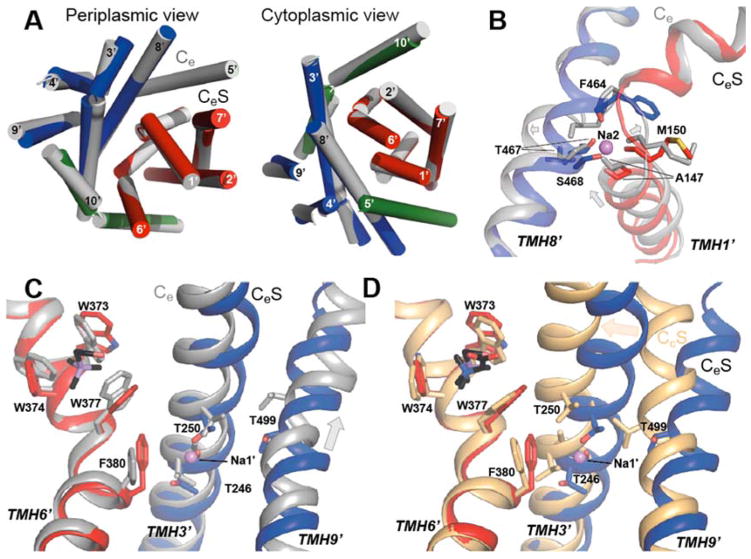
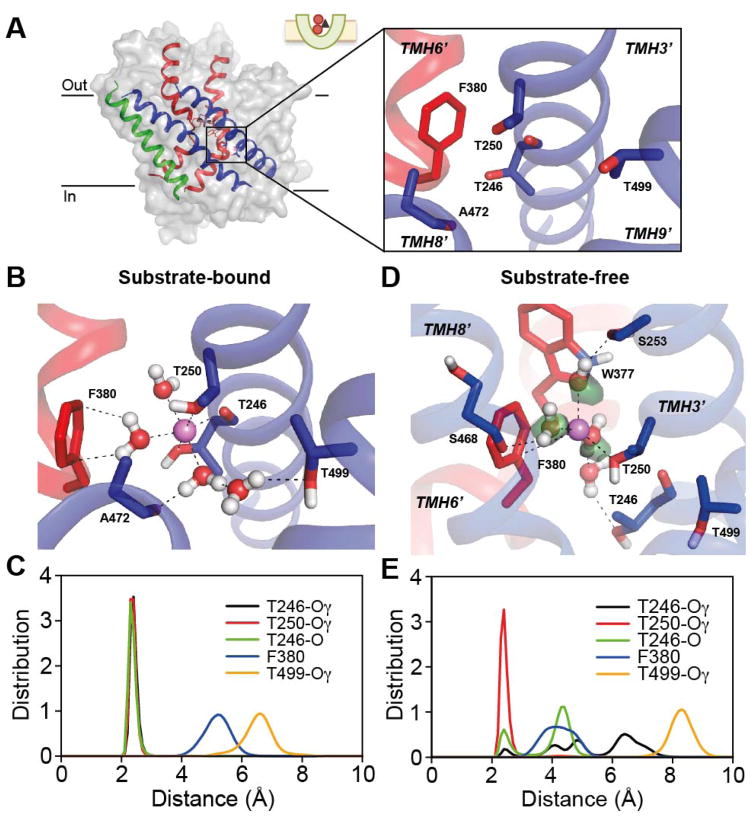
The Na(+)-coupled betaine symporter BetP shares a highly conserved fold with other sequence unrelated secondary transporters, for example, with neurotransmitter symporters. Recently, we obtained atomic structures of BetP in distinct conformational states, which elucidated parts of its alternating-access mechanism. Here, we report a structure of BetP in a new outward-open state in complex with an anomalous scattering substrate, adding a fundamental piece to an unprecedented set of structural snapshots for a secondary transporter. In combination with molecular dynamics simulations these structural data highlight important features of the sequential formation of the substrate and sodium-binding sites, in which coordinating water molecules play a crucial role. We observe a strictly interdependent binding of betaine and sodium ions during the coupling process. All three sites undergo progressive reshaping and dehydration during the alternating-access cycle, with the most optimal coordination of all substrates found in the closed state.


Figures

Figure 1
Figure 1
Binding and transport properties of arseno-choline. Closed squares (choline), open squares (arseno-choline). (A) Tryptophan fluorescence binding curve of choline and arseno-choline to BetP-G153D reconstituted in proteoliposomes. Each point shows the average for eight individual measurements. The error bars represent standard deviation (s.d.). (B) Normalized peak currents of sodium coupled transport of choline and arseno-choline to BetP-G153D. The graphs show average values from three individual recordings and the corresponding s.d.
Figure 2
Figure 2
Conformational states and arseno-choline binding observed in BetP-G153D. (A) Surface representation showing the periplasmic cavity. Chains A and B are in substrate bound outward-open state (CeS), while chain C is in substrate-bound inward-open conformation (CiS). (B) Central binding sites of chains A, B and C, respectively. Arseno-choline is shown in black, red and purple sticks. Anomalous difference Fourier maps are shown at 4.0σ, 7.0σ and 4.0σ levels for substrates in chain A, B and C, respectively.
Figure 3
Figure 3
CeS state coordination of choline in the S1 binding site of BetP-G153D. (A) Typical coordination of choline (black sticks) during the MD simulations by TMH1′ and TMH6′ (cartoon helices). Key residues (sticks) and water molecules (ball-and-stick) are shown. Non-covalent interactions are shown as dashed lines. (B) Distances were measured between the N atom of choline and the center of mass of the side chains in the tryptophan box (W377, W374, and W377), or between the hydroxyl oxygen of choline and the N backbone atom of G151 or the closest carboxyl oxygen atom of D153. The distribution was calculated over three trajectories, each 200 ns long.
Figure 4
Figure 4
The Na2 binding site in outward-open states of BetP-G153D. (A) The CeS state structure (protomer B) in side view with Na2 and adjacent central substrate binding site in stick representation, coordinating Na+ (violet sphere) and arseno-choline (black, red and purple sticks), respectively. The inset shows the coordination of a Na+ ion (violet sphere) in the Na2 site. The FoFc electron density map is shown in green at a level of 3.0σ. (B–E) The Na2 site in simulations of the substrate-bound CeS (B–C) and substrate-free Ce (D–E) states: (B, D) Example configurations in simulations of the CeS state (B) and the Ce state (D). Proteins and ions are presented as in (A), waters are shown as balls-and-sticks and water densities are shown as green surfaces. (C, E) Distance between Na+ ion and Na2-site oxygen atoms during three molecular dynamics simulations of the substrate bound CeS state (C), each 200ns long, and the substrate-free Ce state (E), each 100ns-long. (F) Betaine uptake rates in nmol·min−1·mg−1 cell dry weight (cdw) were measured as a function of the external sodium concentration in E. coli MKH13 cells expressing BetP WT or the mutant N309A. Each point shows the average of at least three independent experiments. Error bars represent s.d.
Figure 5
Figure 5
Comparison of structures and ion binding sites in CeS (colors), Ce (grey) and CcS (tan) states of BetP and BetP-G153D. Sodium ions (violet spheres), and substrate (black sticks) are shown. Changes from the CeS state to the compared state are indicated using arrows. (A–C) Comparisons of substrate-free Ce (PDB entry 4DOJ chain B, grey) and substrate-bound CeS states (colors), showing the effect of substrate binding on the outward-open states on: (A) the overall structure, with helices shown as cylinders; (B) the Na2 binding site, showing protein (ribbons), binding site residues (sticks) and Na2 ion from the CeS structure (sphere). (C) the substrate site (S1) and Na1′ site, with protein shown as ribbons, and with substrate (black) and binding site residues as sticks. The location of the Na1′ ion is taken from the CeS simulations. (D) Comparison of Na1′ and substrate (S1) binding site regions in the substrate-bound outward-open (CeS, colors) and the substrate-bound closed (CcS; PDB entry 4AIN chain B, tan) states. The Na1′ ion location is taken from the CeS simulations.
Figure 6
Figure 6
The Na1′ binding site in outward-open states of BetP-G153D. (A) The CeS state structure (protomer B) in side view with Na1′ site and adjacent central substrate binding site in stick representation, coordinating arseno-choline (black, red and purple sticks). The inset shows residues that form the Na1′ site as reported for the closed substrate-bound CcS state. (B–E) The Na1′ site in simulations of the substrate-bound CeS (B–C) and substrate-free Ce (DE) states: (B, D) Example configurations in simulations of the substrate-bound CeS state (B) and the substrate-free Ce state (D). Proteins and ions are presented as in Fig. 6. (C, E) Distances between the Na+ ion and protein binding site atoms (either oxygen, or the center of the phenyl ring in F380) during simulations of the substrate bound CeS state (C) and the substrate-free Ce state (E).
Figure 7
Figure 7
Comparison of ion binding site accessibility and coordination in the substrate-free Ce (A) and substrate-bound CeS (B) states of BetP-G153D observed using molecular dynamics simulations. The green surface indicates the density of water molecules occupying the extracellular pathway (defined as waters within 16 Å of D153) during three 200 ns trajectories. Protein snapshots were taken at 81 and 83 ns of the trajectories of the Ce and CeS states respectively. BetP protein is shown as a cut-away surface. Sodium ions are shown as violet spheres. Oxygen atoms of water and protein that form the Na2 and Na1′ binding site in the closed CcS state are shown as red spheres. Oxygen atoms in addition to those from the closed state structure coordinating the ion during molecular simulations of the corresponding state are shown as yellow spheres. Selected helices are shown as cartoons: TMH1′ and its symmetry equivalent TMH6′ (red), and TMH3′ and its symmetry equivalent TMH8′ (blue), with substrate choline shown as sticks.
Figure 8
Figure 8
The eight conformations of BetP and BetP-G153D determined by X-ray crystallography to date (green): substrate-free outward-open bound to sodium (Ce2Na) (PDB entry 4DOJ); substrate-bound outward-open (CeS) (PDB entry 4LLH, reported here); substrate-bound closed (CcS) (PDB entry 4AIN); substrate-bound inward-occluded (CiocS) (PDB entry 2WIT); substrate-bound inward-open (CiS) (PDB entries 4DOJ/3P03/4AIN/4LLH); substrate-free inward-open (Ci) (PDB entry 4AMR); substrate-free closed (Cc) (PDB entry 4AIN); and substrate-free outward-occluded (Ceoc) (PDB entry 4DOJ).

References

    1. Yamashita A, Singh SK, Kawate T, Jin Y, Gouaux E. Crystal structure of a bacterial homologue of Na+/Cl−-dependent neurotransmitter transporters. Nature. 2005;437:215–223. - PubMed
    1. Perez C, Koshy C, Yildiz O, Ziegler C. Alternating-access mechanism in conformationally asymmetric trimers of the betaine transporter BetP. Nature. 2012;490:126–130. - PubMed
    1. Forrest LR, Kramer R, Ziegler C. The structural basis of secondary active transport mechanisms. Biochim Biophys Acta. 2011;1807:167–188. - PubMed
    1. Forrest LR, et al. Mechanism for alternating access in neurotransmitter transporters. Proc Natl Acad Sci U S A. 2008;105:10338–10343. - PMC - PubMed
    1. Wong FH, et al. The Amino Acid-Polyamine-Organocation Superfamily. Journal of Molecular Microbiology and Biotechnology. 2012;22:105–113. - PubMed

Publication types

MeSH terms

Substances

Associated data

LinkOut - more resources