. 2014 Jul 21;206(2):183-97.
doi: 10.1083/jcb.201311063. Epub 2014 Jul 14.

DNA damage-specific deubiquitination regulates Rad18 functions to suppress mutagenesis

Affiliations

DNA damage-specific deubiquitination regulates Rad18 functions to suppress mutagenesis

Michelle K Zeman et al. J Cell Biol. .

Abstract

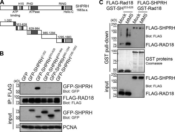
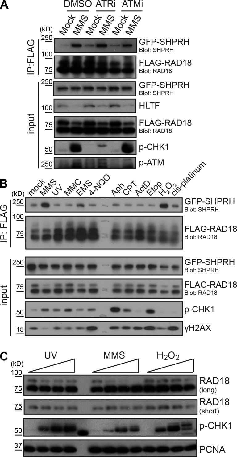
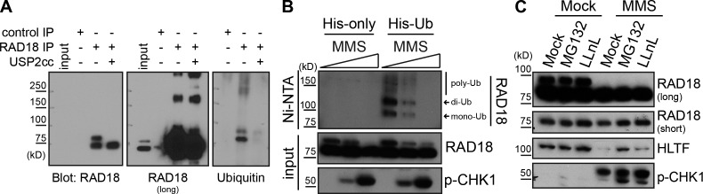
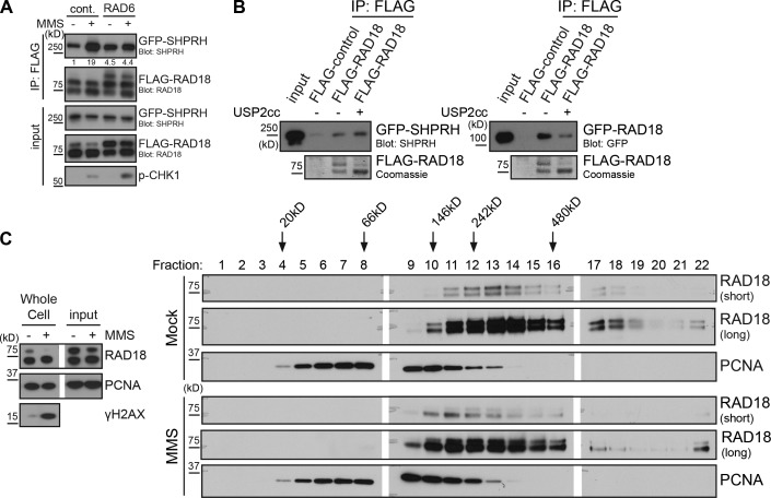
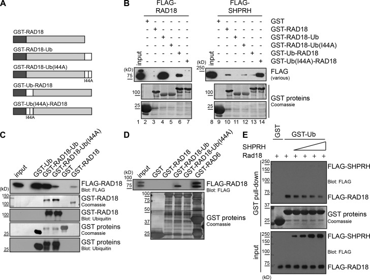
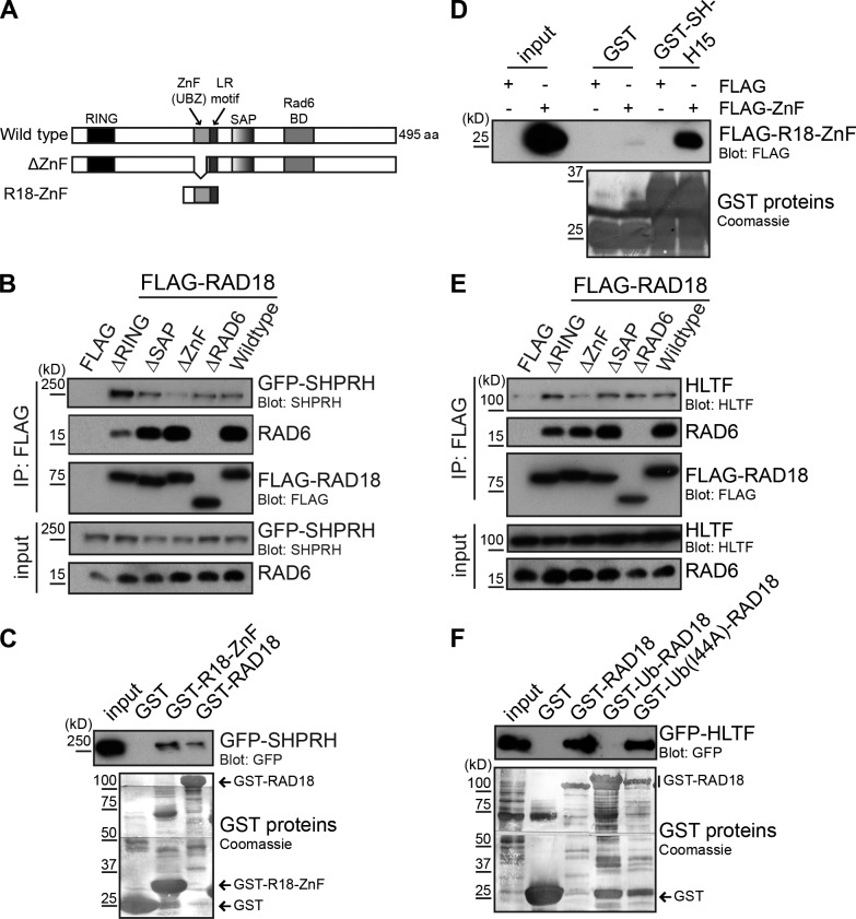
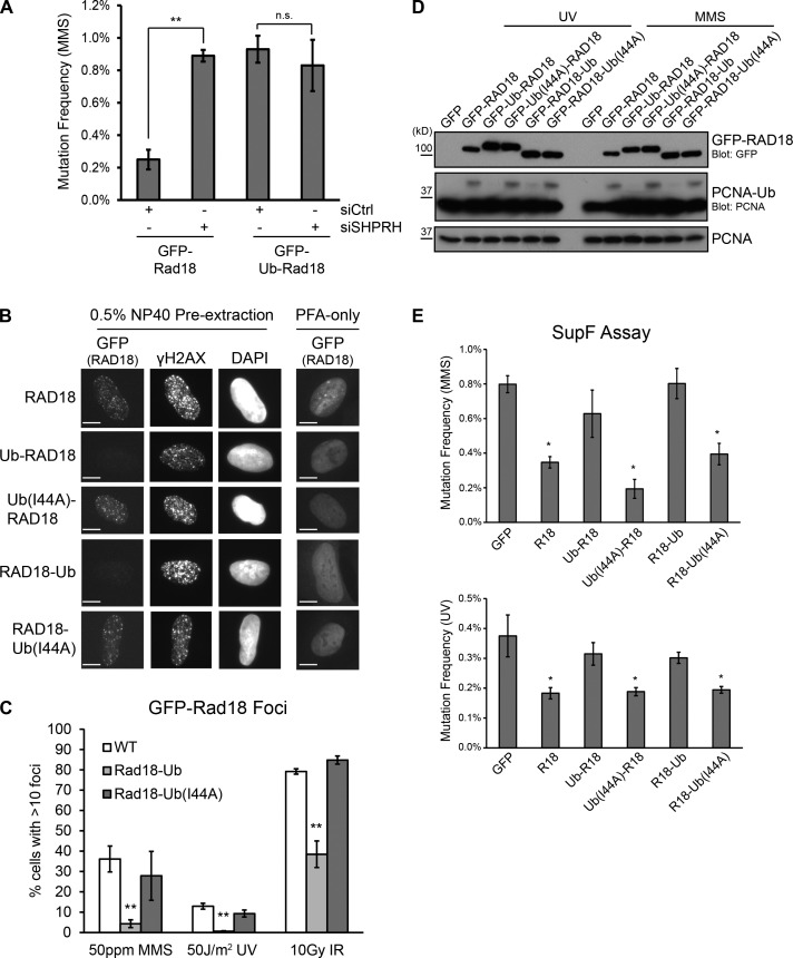
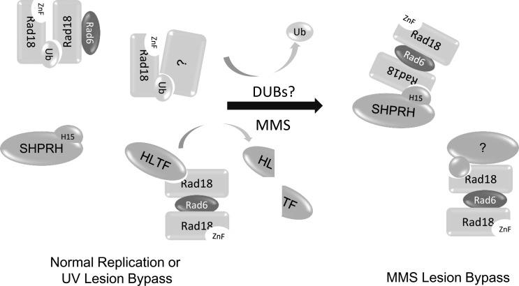
Deoxyribonucleic acid (DNA) lesions encountered during replication are often bypassed using DNA damage tolerance (DDT) pathways to avoid prolonged fork stalling and allow for completion of DNA replication. Rad18 is a central E3 ubiquitin ligase in DDT, which exists in a monoubiquitinated (Rad18•Ub) and nonubiquitinated form in human cells. We find that Rad18 is deubiquitinated when cells are treated with methyl methanesulfonate or hydrogen peroxide. The ubiquitinated form of Rad18 does not interact with SNF2 histone linker plant homeodomain RING helicase (SHPRH) or helicase-like transcription factor, two downstream E3 ligases needed to carry out error-free bypass of DNA lesions. Instead, it interacts preferentially with the zinc finger domain of another, nonubiquitinated Rad18 and may inhibit Rad18 function in trans. Ubiquitination also prevents Rad18 from localizing to sites of DNA damage, inducing proliferating cell nuclear antigen monoubiquitination, and suppressing mutagenesis. These data reveal a new role for monoubiquitination in controlling Rad18 function and suggest that damage-specific deubiquitination promotes a switch from Rad18•Ub-Rad18 complexes to the Rad18-SHPRH complexes necessary for error-free lesion bypass in cells.


Figures

Figure 1.
Figure 1.
MMS-induced modification of Rad18 promotes its interaction with SHPRH. (A) Domain structure of SHPRH. Helic-C, helicase C-terminal domain; PHD, plant homeodomain. (B) The SHPRH353–628 (SH353–628) fragment of SHPRH interacts with Rad18. GFP-tagged SHPRH fragments shown in A were expressed in cells with FLAG-tagged Rad18 and lysed under condition A. FLAG-Rad18 and interacting proteins were analyzed by Western blotting. (C) MMS alters Rad18, not SHPRH, to promote the Rad18–SHPRH interaction. Purified GST-tagged Rad18 or SHPRH353–628 were used to pull down full-length FLAG-SHPRH or FLAG-Rad18 (respectively) from transfected cell lysates, which were mock treated or exposed to 0.005% MMS for 4 h. Associated proteins were analyzed by Western blotting.
Figure 2.
Figure 2.
Damage-inducible Rad18–SHPRH binding coincides with loss of high molecular weight Rad18. (A) Inhibition of checkpoint kinases does not affect the Rad18–SHPRH interaction. Cells transfected with FLAG-Rad18 and GFP-SHPRH were mock treated or exposed to 0.005% MMS and the respective kinase inhibitors for 4 h before being lysed and analyzed as in Fig. 1 B. ATRi, ATR inhibition; ATMi, ATM inhibition. (B) Rad18 and SHPRH interact specifically after MMS or H2O2 treatment. Cells expressing FLAG-Rad18 and GFP-SHPRH were treated with 50 ppm MMS, 50 J/m2 UV, 30 µM mitomycin C (MMC), 0.1% ethyl methanesulfonate (EMS), 20 µM 4-NQO, 30 µM aphidicolin (Aph), 2 µM camptothecin (CPT), 1 µM actinomycin D (ActD), 20 µM etoposide (Etop), 0.1% hydrogen peroxide (H2O2), or 50 µM cis-platinum for 4 h before being lysed under condition B and analyzed by Western blotting. (C) Endogenous Rad18 is deubiquitinated after MMS and H2O2 treatment. Untransfected cells were treated with UV (0, 100, 200, and 400 J/m2), MMS (0, 25, 50, and 100 ppm), or H2O2 (0, 1, 2, and 4 mM) for 4 h before being lysed and analyzed by Western blotting.
Figure 3.
Figure 3.
Rad18 is deubiquitinated after MMS treatment. (A) Endogenous Rad18 is monoubiquitinated. Rad18 was immunoprecipitated (RF antibody) from untransfected cells, and purified protein on beads was treated with or without the Usp2 catalytic core (USP2cc) before being analyzed by Western blotting for the presence of ubiquitin. (B) Rad18 is deubiquitinated after MMS treatment. Cells mock transfected or transfected with His-tagged ubiquitin were treated with MMS (0, 25, and 50 ppm) for 4 h before being lysed under denaturing conditions. His-ubiquitin (Ub) and covalently bound proteins were purified on nickel agarose beads (NTA, nitrilotriacetic acid) and analyzed by Western blotting. (C) Proteasome inhibition does not prevent Rad18 deubiquitination after MMS. Untransfected cells were mock or MMS (0.005%) treated in conjunction with 50 µM MG132 or 25 µM LLnL for 4 h and processed as in Fig. 2 C.
Figure 4.
Figure 4.
Direct modulation of Rad18 ubiquitination affects Rad18–Rad18 and Rad18–SHPRH interactions. (A) Promoting Rad18 ubiquitination in vivo prevents damage-inducible binding to SHPRH. Cells were cotransfected with GFP-SHPRH, FLAG-Rad18, and either Rad6 or empty vector and then mock or MMS (0.005%) treated for 4 h before being lysed and processed as in Fig. 2 B. Quantification indicates ratio of GFP-SHPRH in the IP relative to the respective input sample, normalized to the mock-transfected, mock-treated control (cont.). (B) Deubiquitinating Rad18 affects its protein–protein interactions. FLAG-Rad18 was cotransfected with Rad6 and then purified from lysates under high-salt conditions. Beads were mock treated or deubiquitinated by Usp2 and used to pull-down GFP-SHPRH or GFP-Rad18 from transfected cells lysed as in Fig. 1 C. Pull-downs were analyzed by Western blotting. USP2cc, Usp2 catalytic core. (C) Rad18 shifts to a smaller complex after MMS treatment. Cells were mock treated or exposed to 0.01% MMS for 2 h and then lysed and separated on a 5–30% glycerol gradient. Gradient fractions were analyzed for the presence of Rad18 and PCNA by Western blotting. White lines indicate that intervening lanes have been spliced out.
Figure 5.
Figure 5.
Rad18-Ub fusion proteins preferentially bind to nonubiquitinated Rad18. (A) Structure of GST–Rad18-Ub chimera proteins. GST (dark gray), ubiquitin (white), and Rad18 (light gray) were fused in frame. The I44A mutation in ubiquitin prevents recognition by a ZnF ubiquitin-binding domain (Bomar et al., 2007). (B) Fusing ubiquitin to Rad18 promotes binding to Rad18 but not to SHPRH. GST-Rad18 fusion proteins were incubated with whole-cell lysates transfected with FLAG-Rad18 or FLAG-SHPRH and analyzed as in Fig. 1 C. (C) The Rad18–Rad18 interaction is weaker than the Rad18-Ub–Rad18 interaction. GST-Rad18 fusion proteins were incubated with whole-cell lysates transfected with FLAG-Rad18 and analyzed as in Fig. 1 C. Ubiquitin blots were used to confirm the identity of the GST constructs. (D) Rad18-Ub chimeras bind preferentially to nonubiquitinated Rad18. GST-Rad18 fusion proteins were incubated with cell lysates cotransfected with FLAG-Rad18 and Rad6 to attain a ratio of ∼1:1 Rad18/Rad18•Ub, and analyzed as in Fig. 1 C. (E) SHPRH and ubiquitin compete for binding to Rad18. GST-tagged ubiquitin on beads was incubated with FLAG-Rad18 and increasing amounts of FLAG-SHPRH, both purified from human cells. Proteins bound to the beads were analyzed by Western blotting.
Figure 6.
Figure 6.
Rad18 binds SHPRH and HLTF through its ubiquitin-binding ZnF domain. (A) Domain structure of Rad18 showing wild-type, Rad18 ΔZnF(Δ200–224), and Rad18-ZnF(186–240) constructs (Huang et al., 2009). BD, binding domain. (B) The Rad18-ZnF is important to bind SHPRH. FLAG-Rad18 constructs lacking the indicated domains were cotransfected with GFP-SHPRH. FLAG-Rad18 and interacting proteins were analyzed as in Fig. 1 B. (C) The Rad18-ZnF contributes to SHPRH binding. GST–Rad18-ZnF or full-length Rad18 was used to pull-down GFP-SHPRH from lysates and analyzed as in Fig. 1 C. (D) The Rad18-ZnF interacts directly with the SHPRH-H15 domain. GST–SHPRH-H15 was used to pull-down FLAG–Rad18-ZnF from cell lysates and analyzed as in Fig. 1 C. (E) The Rad18-ZnF is important for binding HLTF. FLAG-Rad18 constructs lacking the indicated domains were cotransfected with untagged HLTF. FLAG-Rad18 and interacting proteins were analyzed as in Fig. 1 B. (F) HLTF binding is disrupted by the Rad18-Ub fusion. GST-Rad18 fusion proteins were incubated with cell lysates transfected with GFP-HLTF and analyzed as in Fig. 1 C.
Figure 7.
Figure 7.
Rad18-Ub fusions are unable to respond to DNA damage. (A) Knockdown of SHPRH and overexpression of Rad18-Ub is epistatic. SupF reporter plasmid (0.5% MMS) was recovered from HEK 293T cells 48 h after cotransfection with the indicated GFP-Rad18 constructs (Ub, ubiquitin) and siRNAs (siCtrl, control siRNA). Data represent mean and standard deviations from three independent experiments. At least 2,000 colonies were analyzed per condition. **, P < 0.01, relative to control knockdown. (B) Rad18-Ub chimeras are nuclear but fail to localize to sites of DNA damage. U2OS clones stably expressing different forms of GFP-Rad18 were treated with 0.005% MMS for 4 h before preextraction and fixation. Rad18 was visualized using direct GFP fluorescence, γ-H2AX was detected using immunofluorescence. Bars, 10 µm. (C) Rad18-Ub chimeras do not form foci after any type of DNA damage. Clones in B were treated with MMS for 4 h or allowed to recover for 4 h after exposure to the indicated doses of UV or ionizing radiation (IR). GFP-Rad18 foci were counted using ImageJ. Each bar represents a mean and standard deviation from three independent experiments. At least 100 cells were counted per condition. **, P < 0.01, relative to wild-type (WT) Rad18. (D) Rad18-Ub chimeras cannot induce PCNA ubiquitination. Rad18−/− HCT116 cells were transiently transfected with GFP-Rad18 chimera constructs and damaged with UV or MMS before being analyzed as in Fig. 2 C. (E) Rad18-Ub chimeras cannot suppress mutagenesis on MMS- or UV-damaged plasmids. SupF reporter plasmid (0.5% MMS or 1,000 J/m2 UV) was recovered from Rad18−/− HCT116 cells 48 h after cotransfection with the indicated GFP-Rad18 chimera constructs (Ub(I44A), ubiquitin-I44A). Data represent means and SEMs from four independent experiments. At least 2,000 colonies were analyzed per condition. *, P < 0.05, relative to GFP-only control. R18, Rad18.
Figure 8.
Figure 8.
Ubiquitination of Rad18 controls its interactions and functions. In undamaged cells, Rad18 exists in equilibrium between the ubiquitinated and nonubiquitinated state and forms complexes with distinct binding partners. Upon treatment with MMS, HLTF is degraded, and Rad18 is deubiquitinated, freeing the ZnF domain of Rad18 to interact with the H15 domain of SHPRH and inducing formation of SHPRH–Rad18 complexes, which prevents MMS-induced mutagenesis.

References

    1. Achar, Y.J., Balogh D., and Haracska L.. 2011. Coordinated protein and DNA remodeling by human HLTF on stalled replication fork. Proc. Natl. Acad. Sci. USA. 108:14073–14078 10.1073/pnas.1101951108 - DOI - PMC - PubMed
    1. Bi, X., Barkley L.R., Slater D.M., Tateishi S., Yamaizumi M., Ohmori H., and Vaziri C.. 2006. Rad18 regulates DNA polymerase κ and is required for recovery from S-phase checkpoint-mediated arrest. Mol. Cell. Biol. 26:3527–3540 10.1128/MCB.26.9.3527-3540.2006 - DOI - PMC - PubMed
    1. Bienko, M., Green C.M., Sabbioneda S., Crosetto N., Matic I., Hibbert R.G., Begovic T., Niimi A., Mann M., Lehmann A.R., and Dikic I.. 2010. Regulation of translesion synthesis DNA polymerase η by monoubiquitination. Mol. Cell. 37:396–407 10.1016/j.molcel.2009.12.039 - DOI - PubMed
    1. Bish, R.A., and Myers M.P.. 2007. Werner helicase-interacting protein 1 binds polyubiquitin via its zinc finger domain. J. Biol. Chem. 282:23184–23193 10.1074/jbc.M701042200 - DOI - PubMed
    1. Blastyák, A., Hajdú I., Unk I., and Haracska L.. 2010. Role of double-stranded DNA translocase activity of human HLTF in replication of damaged DNA. Mol. Cell. Biol. 30:684–693 10.1128/MCB.00863-09 - DOI - PMC - PubMed

Publication types