Review
. 2014;58(2-4):219-29.
doi: 10.1387/ijdb.140109ss.

Natural killer cells and regulatory T cells in early pregnancy loss

Affiliations
Review

Natural killer cells and regulatory T cells in early pregnancy loss

Surendra Sharma. Int J Dev Biol. 2014.

Abstract

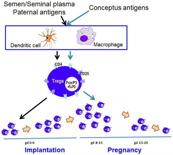
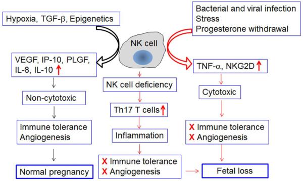
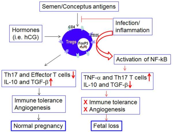
Survival of the allogeneic embryo in the uterus depends on the maintenance of immune tolerance at the maternal-fetal interface. The pregnant uterus is replete with activated maternal immune cells. How this immune tolerance is acquired and maintained has been a topic of intense investigation. The key immune cells that predominantly populate the pregnant uterus are natural killer (NK) cells. In normal pregnancy, these cells are not killers, but rather provide a microenvironment that is pregnancy compatible and supports healthy placentation. In placental mammals, an array of highly orchestrated immune elements to support successful pregnancy outcome has been incorporated. This includes active cooperation between maternal immune cells, particularly NK cells, and trophoblast cells. This intricate process is required for placentation, immune regulation and to remodel the blood supply to the fetus. During the past decade, various types of maternal immune cells have been thought to be involved in cross-talk with trophoblasts and in programming immune tolerance. Regulatory T cells (Tregs) have attracted a great deal of attention in promoting implantation and immune tolerance beyond implantation. However, what has not been fully addressed is how this immune-trophoblast axis breaks down during adverse pregnancy outcomes, particularly early pregnancy loss, and in response to unscheduled inflammation. Intense research efforts have begun to shed light on the roles of NK cells and Tregs in early pregnancy loss, although much remains to be unraveled in order to fully characterize the mechanisms underlying their detrimental activity. An increased understanding of host-environment interactions that lead to the cytotoxic phenotype of these otherwise pregnancy compatible maternal immune cells is important for prediction, prevention and treatment of pregnancy maladies, particularly recurrent pregnancy loss. In this review, we discuss relevant information from experimental and human models that may explain the pregnancy disrupting roles of these pivotal sentinel cells at the maternal-fetal interface.


Figures

Fig. 1
Fig. 1. Generation of semen/seminal fluid and conceptus-specific uterine regulatory T cells (Tregs) in the mouse
During the pre-implantation phase, Tregs can be generated in response to semen/seminal plasma paternal antigens. These cells have been shown to play an important role in implantation. However, Tregs reach their peak around gestational day 12 or 13. We propose that conceptus-specific Tregs contribute to influx of CD4+CD25+FoxP3+ Tregs after implantation. These Tregs could be of extrathymic origin and support maintenance of immune tolerance and promote angiogenesis. These cells start tapering off after gestational day 13 and reach basal numbers at the end of pregnancy. It is possible that Tregs could also be modified or poorly propagated in response to hormone dyregulation and/or inflammatory environment.
Fig. 2
Fig. 2. Can uterine NK (uNK) cells become detrimental to normal pregnancy?
In humans, non-cytotoxic uNK cells are phenotypically characterized as CD56brightCD16 and express full spectrum of natural cytotoxicity receptors (NCRs) and cytotoxic machinery. Their mouse counterparts are typically shown as DBA+ and may lose expression of NK1.1 and DX5 markers. Recent observations suggest that in both cases, uNK cells are potent producers of angiogenic factors and anti-inflammatory molecules. These properties make them helpers, rather than killers, of pregnancy. This pregnancy compatible role of uNK cells has been shown to be compromised in the scenarios of NK cell deficiency and exposure to inflammatory triggers. NK cell deficiency has been shown to encourage heavy recruitment of Th17 T cells which cause pathogenic inflammation. Exposure to inflammatory triggers such as bacterial and viral mimics (LPS or poly I:C) may either convert uNK cells into TNF-α-producing cells or encourage recruitment of NK cells which become TNF-α producing and NKG2Dbright uNK cells. In an environment of poor immune tolerance and impaired angiogenesis, these NK cells are likely to induce fetal loss.
Fig. 3
Fig. 3. Acquisition of inflammatory phenotype by uterine regulatory T cells (Tregs)
As proposed in Figure 1, two sets of uterine Tregs could be generated during pregnancy. Semen/seminal fluid specific Tregs develop early and populate the implantation site. Their depletion or their poor development and recruitment may lead to implantation failure. Importantly, we propose that uterine Tregs could also be converted into inflammatory Tregs in response to bacterial and viral triggers. This could also lead to excessive production of TNF-α and presence of Th17 T cells. This pathway will be the cause of break-down in immune tolerance and angiogenesis and eventually lead to fetal loss. Tregs are identified as cells intrinsically expressing surface T cell markers CD4 and CD25 and the nuclear transcription factor FoxP3.

References

    1. ALUVIHARE VR, KALLIKOURDIS M, BETZ AG. Regulatory T cells mediate maternal tolerance to the fetus. Nat Immunol. 2004;5:266–271. - PubMed
    1. ARNON TI, MARKEL G, MANDELBOIM O. Tumor and viral recognition by natural killer cells receptors. Sem Cancer Biol. 2006;16:348–358. - PubMed
    1. ARPAIA N, CAMPBELL C, FAN X, DIKIY S, VAN DER VEEKEN J, DEROOS P, LIU H, CROSS JR, PFEFFER K, COFFER PJ, RUDENSKY AY. Metabolites produced by commensal bacteria promote peripheral regulatory T-cell generation. Nature. 2013;504:451–455. - PMC - PubMed
    1. ASHKAR AA, CROY BA. Interferon-gamma contributes to the normalcy of murine pregnancy. Biol Reprod. 1999;61:493–502. - PubMed
    1. BALANDYA E, WIELAND-ALTER W, SANDERS K, LAHEY T. Human seminal plasma fosters CD4(+) regulatory T cell phenotype and transforming growth factor –b1 expression. Am J Reprod Immunol. 2012;68:322–330. - PMC - PubMed

Publication types