. 2014 Jul 15:5:4393.
doi: 10.1038/ncomms5393.

Cholesterol selectively activates canonical Wnt signalling over non-canonical Wnt signalling

Affiliations

Cholesterol selectively activates canonical Wnt signalling over non-canonical Wnt signalling

Ren Sheng et al. Nat Commun. .

Abstract

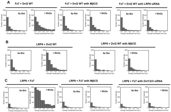
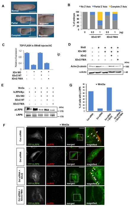
Wnt proteins control diverse biological processes through β-catenin-dependent canonical signalling and β-catenin-independent non-canonical signalling. The mechanisms by which these signalling pathways are differentially triggered and controlled are not fully understood. Dishevelled (Dvl) is a scaffold protein that serves as the branch point of these pathways. Here, we show that cholesterol selectively activates canonical Wnt signalling over non-canonical signalling under physiological conditions by specifically facilitating the membrane recruitment of the PDZ domain of Dvl and its interaction with other proteins. Single-molecule imaging analysis shows that cholesterol is enriched around the Wnt-activated Frizzled and low-density lipoprotein receptor-related protein 5/6 receptors and plays an essential role for Dvl-mediated formation and maintenance of the canonical Wnt signalling complex. Collectively, our results suggest a new regulatory role of cholesterol in Wnt signalling and a potential link between cellular cholesterol levels and the balance between canonical and non-canonical Wnt signalling activities.


Figures

Fig. 1
Fig. 1. Cholesterol binding of Dvl2 PDZ domain
(A) Cholesterol dependence of membrane binding of Dvl2-PDZ. Binding of the PDZ domain to POPC/POPS/cholesterol (80-x:20:x, x = 0-40 mole%) vesicles was measured by SPR analysis. Notice that Dvl2-PDZ has significant binding to POPC/POPS (80:20) vesicles and thus the net affinity for cholesterol should be estimated by subtracting the sensorgram for POPC/POPS (80:20) vesicles from that of particular vesicles. Equilibrium SPR measurements were performed in 20 mM Tris-HCl, pH 7.4, containing 0.16 M KCl. POPC vesicles to which Dvl2-PDZ has no detectable affinity were used for the control surface. (B) A model structure of the Dvl2-PDZ-cholesterol-peptide ternary complex was obtained by docking cholesterol and the C-terminal Fz7 peptide to Dvl2-PDZ (PDB ID: 2REY). Cholesterol (with its hydrophobic tail pointing upward) and the bound peptide are shown in space-filling and stick representations, respectively. Putative cholesterol-binding residues, F227 and M300, are shown in stick representation and labeled. They are predicted to partially penetrate the membrane and make contact with the hydrophobic A and B rings of cholesterol. The structure on the left panel is shown in a putative membrane-binding orientation with the dotted line indicating the membrane surface. The same model structure is also shown in a different orientation with the top surface facing the membrane (right panel). Notice that the putative lipid binding sites are topologically distinct from the peptide-binding pocket. The model was built in the absence of the lipid bilayer and the protein may undergo further conformational changes when it binds the membrane.
Fig. 2
Fig. 2. Single molecule tracking of Dvl and other members of the canonical Wnt signaling complex
(A) A representative pair of EGFP-tagged Dvl2 (green circles) and Halo-TMR-labeled Fz7 (red circles) in a HeLa cell is shown. The left panel is for Dvl2 WT and the right panel for FM/A. Many Dvl2 WT molecules show dynamic colocalization with Fz7 in the presence of Wnt3a (>5 minutes after Wnt3a stimulation) whereas most of FM/A molecules exhibit separate localization with Fz7 (see also Supplementary Movie 2 and 4). Green and red dots indicate individual Dvl2 and Fz7 molecules, respectively, and lines are their trajectories. Yellow scale bars indicate 0.5 μm. (B) Distribution of colocalization time of Halo-TMR-Fz7 with EGFP-Dvl2 WT or FM/A. The number of Fz7 molecules spending a given colocalzation time with Dvl2 WT on the PM of HeLa cells is displayed. Notice that Wnt3a dramatically enhanced the population of Fz7 molecules co-localized with Dvl2 WT for >100 msec. In contrast, no Fz7 molecule was found to colocalized with the Dvl2 FM/A mutant for >100 msec with or without Wnt3a (see also Supplementary Movie 1-4). Wnt5a had no effect on Dvl2 WT or FM/A. The same size of PM surface was analyzed for each histogram. (C) Distribution of colocalization time of EGFP-LRP6 with Halo-TMR-Dvl2 WT or FM/A. Wnt3a drastically enhanced the co-localization of Dvl2-WT (not FM/A) and LRP6 whereas Wnt5a had no effect on Dvl2 WT or FM/A (D) Distribution of colocalization time of EGFP-Axin with Halo-TMR-Dvl2 WT or FM/A. Wnt3a treatment significantly increased the population of co-localized Dvl2 WT-Axin pairs while having no effect on Dvl2 FM/A-Axin co-localization. Wnt5a showed no detectable effect. (E) Distribution of colocalization time of Halo-TMR-Fz7 with EGFP-Dvl2-Δ424-507 (or Δ424-507-FM/A). The two truncation mutants behaved essentially the same as their non-truncated counterparts shown in Fig 2B. 50 ng/ml Wnt3a or Wnt5a was used in all experiments.
Fig. 3
Fig. 3. Effects of cholesterol depletion and protein knockdown on interactions among Wnt signaling proteins in HeLa cells
(A) Effects of cholesterol depletion by methyl-β-cyclodextrin (MβCD) and LRP6 knockdown on Dvl2-Fz7 co-localization before and 10 minutes after stimulation by Wnt3a. (B) Effect of cholesterol depletion by MβCD on Dvl2-LRP6 co-localization before and 10 minutes after stimulation by Wnt3a. (C) Effects of cholesterol depletion by MβCD and Dvl1/2/3 triple knockdown on Dvl2-Fz7 co-localization before and 10 minutes after stimulation by Wnt3a. Protein co-localization was determined by single molecule imaging as described for Fig. 2. 50 ng/ml Wnt3a was used for all experiments. Cholesterol depletion was achieved by incubating cells with 1 mM MβCD in DMEM for 1 hour at 37oC. 50 nM siRNA was used for each knockdown experiment.
Fig. 4
Fig. 4. Wnt-induced local cholesterol concentration changes estimated by a cholesterol probe
(A) Distribution of colocalization time of Halo-TMR-Fz7 with EGFP-D4-D434A before and after Wnt3a or Wnt5a treatment. Colocalization was dramatically increased in response to Wnt3a stimulation and lasted >20 minutes under our experimental conditions. (B) Distribution of colocalization time of Halo-TMR-D4-D434A with EGFP-LRP6 before and after Wnt3a and Wnt5a treatment. Notice that D4-D434A and EGFP-LRP6 are significantly co-localized even in the absence of Wnt3a; consequently, Wnt3a stimulation enhanced the overall co-localization only modestly. 50 ng/ml Wnt3a or Wnt5a was used in all experiments.
Fig. 5
Fig. 5. Cholesterol binding of Dvl is required for canonical Wnt signaling under physiological conditions
(A) Induction of the secondary axis by exogenous addition of XDvl2. The partial axis was characterized by the axial structure without the head structure including eyes and cement glands. The complete axis has the axial structure along with the head structure. Scale bars indicate 1 mm. (B) Quantification of Fig. 5A. (C) The TOP-FLASH activity was measured in Xenopus AC explants. XWnt8 mRNAs were injected with indicated XDvl2 mRNAs and XDvl Morpholinos (MO). At stage 10, ACs were dissected and analyzed. Error bars indicate standard deviations from three independent analyses. (D) The Western blot analysis of activated (dephosphorylated) β-catenin levels. α-Actin was used as a loading control. (E) Western blot data showing the degree of LRP6 phosphorylation. Xenopus embryos were injected with indicated mRNAs, including human Myc-tagged LRP6 (hLRP6-Myc), and AC tissue explants were analyzed at stage10.5. Only exogenous Dvl2 WT could rescue Dvl MO-mediated inhibition of LRP6 phosphorylation in Xenopus AC tissues. hLRP6-Myc was immunoprecipitated by the c-Myc antibody and phosphorylated LRP6 (pLRP6) was detected with the antibody against Sp1490. (F) Confocal images of HeLa cells showing the location and degree of LRP6 phosphorylation. The Wnt3a-mediated colocalization (see white arrows) of GFP-human LRP6 (hLRP6) (Green) and phosphorylated LRP6 at Ser1490 (pLRP6: Red) was clearly seen at the PM and endosomes in control cells (Con siRNA: first row). Dvl siRNA (second row) abrogated the colocalization of hLRP6 and pLRP6, which was significantly rescued by XDvl2 (third row), but not by XDvl2 FM/A (bottom row). Scale bars indicate 20 μm (G) Quantification of Fig. 5F.
Fig. 6
Fig. 6. Differential Effects of cholesterol on canonical and non-canonical Wnt signaling during Xenopus embryogenesis
(A) Xnr3 expression was analyzed by in situ hybridization. Embryos were injected with the indicated reagents at the dorsal side of 4-cell stage with a lineage tracer β-galactosidase mRNA and analyzed at stage 10.5. The scale bar indicates 250 μm. (B) Quantification of Fig. 6A. (C) One side of each embryo was injected with the indicated reagents at the 4-cell stage with a β-galactosidase mRNA. Expressions of MyoD at stage12, and BF1, En2 and Twist at stage 17-19 were monitored on both injected (inj.) and uninjected (Uninj.) sides by in situ hybridization. The scale bar indicates 250 μm. (D) Quantification of Fig. 6C. Percentage of embryos with reduced gene expression on the injected side was counted. (E) Reagents were introduced into the dorsal marginal region of 4-cell stage embryos and defective phenotypes were counted at tadpole stages. Severe defects were characterized by the shortened and kinked body axis and mild defects by the slightly shortened and defective head structure. Scale bars indicate 1 mm. (F) Quantification of Fig. 6E. (G) DMZs were dissected at stage 10 and cultured up to stage 18. Each image was a representative of three independent experiments. Scale bars indicate 0.4 mm. (H) Quantification of Fig. 6G. The Y-axis indicates the length/width ratio (L/W) of each DMZs. Error bars denote standard deviations. Asterisk indicates p < 0.005 (determined by two-tailed t-test) when compared to the control, (XDvl MO + XDvl2)- and (XDvl MO + XDvl2 FM/A)-injected explants, respectively. For all experiments, endogenous XDvl levels were suppressed by triple MO, and XDvl2 WT or FM/A was reintroduced to embryos.
Fig. 7
Fig. 7. Cholesterol binding of Dvl2 is required only for canonical Wnt signaling in culture cells
(A-C) TOP-Flash activity (A), Activated β-catenin level (B) and phosphorylated JNK level (C) were measured in HEK293FT cells. (A) HEK293FT cells were transfected with 50 nM of control siRNA or Dvl1/2/3 siRNAs along with TOP-Flash reporter constructs and Dvl DNAs prior to 24 hours of Wnt3a conditioned medium (CM) stimulation. Error bars indicate standard deviations from three independent analyses. (B,C) HEK293FT cells were transfected with indicated siRNAs and DNAs prior to 6 hours of Wnt3a stimulation (B) or 1 hour of Wnt11 stimulation (C). Cell lysates were harvested and analyzed by western blotting with activated β-catenin (B) or phosphorylated JNK antibodies (C). Actin and JNK levels were used as loading controls.
Fig 8
Fig 8. A model for cholesterol-mediated canonical Wnt signaling-specific functions of Dvl
(A) Binding of a canonical Wnt to the Fz7 and LRP6 receptors induces the local enrichment of cholesterol in the vicinity of the activated receptor complex by bringing Fz7 closer to LRP6 in cholesterol-enriched microenvironment. This concomitantly recruits Dvl to the complex through its coincident recognition of Fz7 and cholesterol (and an anionic lipid, such as PtdIns(4,5)P2) by its PDZ domain. Cholesterol binding of Dvl ensures the canonical Wnt signaling-specific scaffolding function of Dvl by allowing sustained interaction of Dvl with Fz7 and other proteins involved in canonical Wnt signaling, including Axin and GSK3, and thereby facilitating LRP6 phosphorylation. The canonical Wnt signaling complex containing phosphorylated LRP6 can also be endocytosed to continue signaling activities intracellularly. (B) By contrast, cholesterol binding of Dvl is not required for non-canonical Wnt signaling (e.g., Wnt/PCP signaling) because its scaffolding activity for proteins involved in these pathways (e.g., Daam1) does not seem to depend on its sustained PM localization. Co-R indicates a potential co-receptor for non-canonical Wnt signaling. Other putative steps of canonical Wnt signaling, including receptor complex aggregation, local PtdIns(4,5)P2 enrichment ,,, and the downstream Wnt signaling events are omitted here for simplicity.

References

    1. Logan CY, Nusse R. The Wnt signaling pathway in development and disease. Annu. Rev. Cell Dev. Biol. 2004;20:781–810. - PubMed
    1. Clevers H. Wnt/beta-catenin signaling in development and disease. Cell. 2006;127:469–480. - PubMed
    1. Klaus A, Birchmeier W. Wnt signalling and its impact on development and cancer. Nat. Rev. Cancer. 2008;8:387–398. - PubMed
    1. MacDonald BT, Tamai K, He X. Wnt/beta-catenin signaling: components, mechanisms, and diseases. Dev. Cell. 2009;17:9–26. - PMC - PubMed
    1. Gao C, Chen YG. Dishevelled: The hub of Wnt signaling. Cell. Signal. 2010;22:717–727. - PubMed

Publication types