. 2014 Jul 29;111(30):E3053-61.
doi: 10.1073/pnas.1411082111. Epub 2014 Jul 14.

Platelets guide the formation of early metastatic niches

Affiliations

Platelets guide the formation of early metastatic niches

Myriam Labelle et al. Proc Natl Acad Sci U S A. .

Abstract

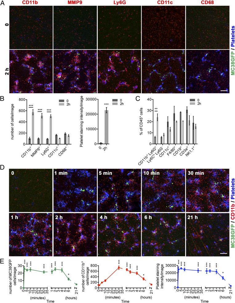
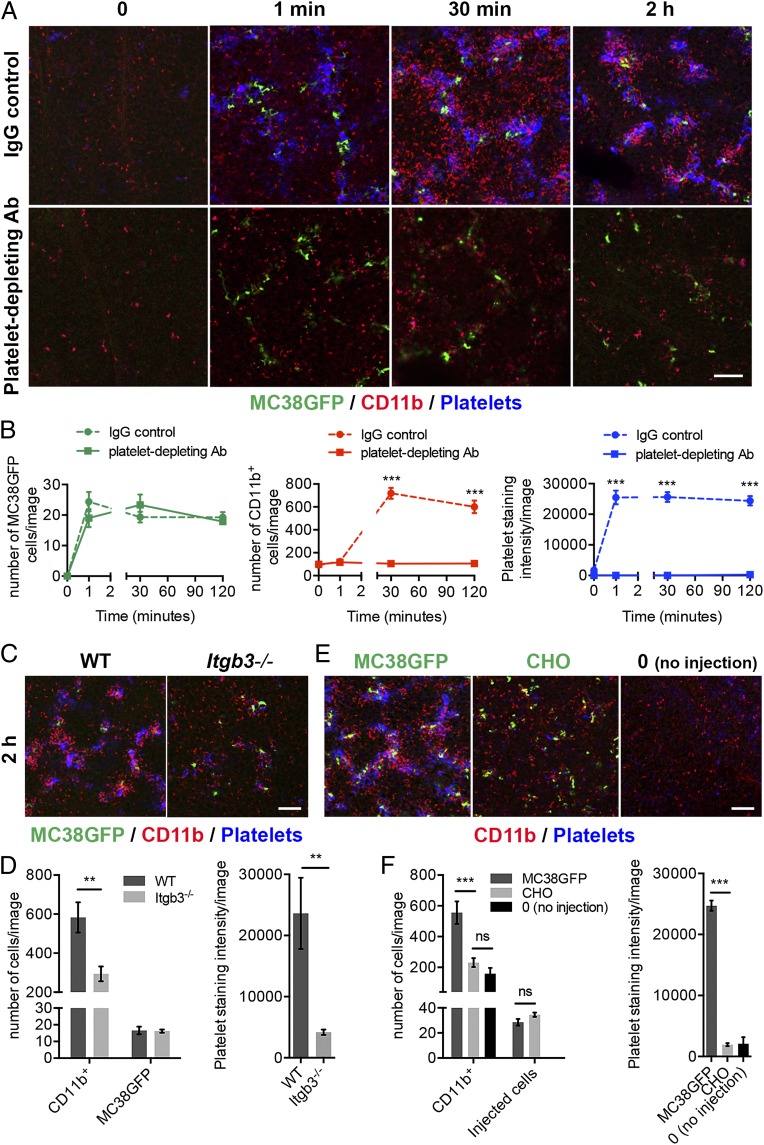
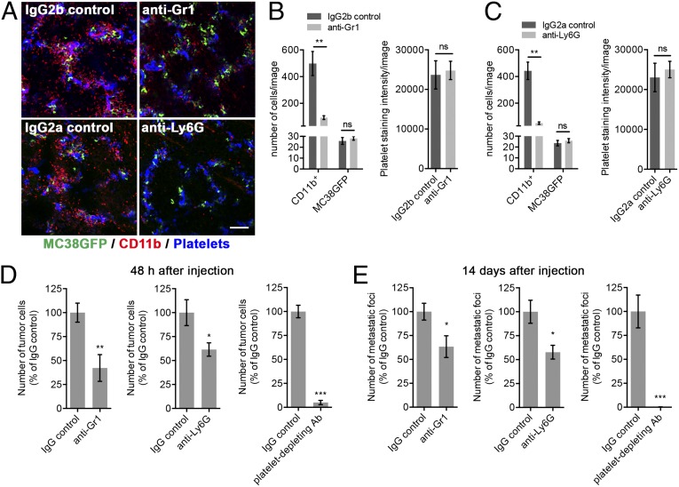
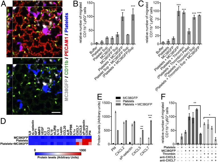
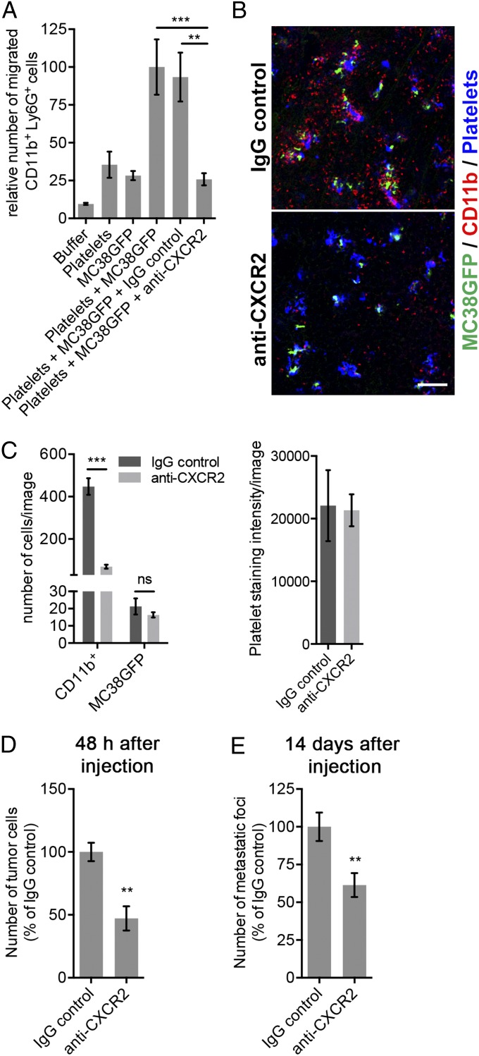
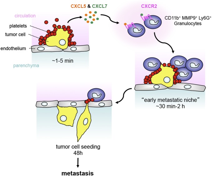
During metastasis, host cells are recruited to disseminated tumor cells to form specialized microenvironments ("niches") that promote metastatic progression, but the mechanisms guiding the assembly of these niches are largely unknown. Tumor cells may autonomously recruit host cells or, alternatively, host cell-to-host cell interactions may guide the formation of these prometastatic microenvironments. Here, we show that platelet-derived rather than tumor cell-derived signals are required for the rapid recruitment of granulocytes to tumor cells to form "early metastatic niches." Granulocyte recruitment relies on the secretion of CXCL5 and CXCL7 chemokines by platelets upon contact with tumor cells. Blockade of the CXCL5/7 receptor CXCR2, or transient depletion of either platelets or granulocytes prevents the formation of early metastatic niches and significantly reduces metastatic seeding and progression. Thus, platelets recruit granulocytes and guide the formation of early metastatic niches, which are crucial for metastasis.


Conflict of interest statement

The authors declare no conflict of interest.

Figures

Fig. 1.
Fig. 1.
CD11b+MMP9+Ly6G+ granulocytes are recruited to platelet–tumor cell aggregates within 2 h of tumor cell entry into the circulation. (A) Immunostaining of lungs from control mice (0; no injection) or from mice killed 2 h after the injection of 106 MC38GFP tumor cells (green). Staining for the immune cell markers CD11b, MMP9, Ly6G, CD11c, or CD68 (red) reveals that CD11b+, MMP9+, and Ly6G+ cells are recruited to the platelet (blue)–tumor cell (green) aggregates, whereas CD11c+ and CD68+ cells are not. Platelets were stained with an anti-GP1bβ antibody (blue). (Scale bar: 100 µm.) See Fig. S1 for CD11b, Ly6G, and MMP9 costaining. (B) Numbers of CD11b+, MMP9+, Ly6G+, CD11c+, and CD68+ cells (Left), and intensity of platelet staining (Right) in lungs of mice treated as in A. Bars represent the mean ± SEM (n ≥ 12 images from n ≥ 3 mice). ***P < 0.001 was determined by unpaired two-sided t test. (C) Mouse lungs were collected at time 0 or 2 h after the injection of 106 MC38GFP tumor cells, and the percentages of different immune cell populations among total immune cells (CD45+) were determined by FACS. Bars represent the mean ± SEM (n ≥ 3 mice). **P < 0.01 was determined by unpaired two-sided t test. (D) Time course of the formation of the early metastatic niche. Immunostaining for CD11b (red), and platelets (GP1bβ; blue) in mouse lungs at the times indicated following the injection of 106 MC38GFP cells (green). Platelet–tumor cell aggregates are formed within 1 min of MC38GFP cell entry into the circulation. In comparison, the recruitment of CD11b+ cells (red) to the platelet–tumor cell aggregates requires 30 min to 4 h. Note the clear enrichment for CD11b+ cells in the vicinity of tumor cells at 2 and 4 h. (Scale bar: 100 µm.) (E) Numbers of MC38GFP tumor cells (Left) and CD11b+ cells (Center), and intensity of platelet staining (Right) in mouse lungs collected at the times indicated following the injection of MC38GFP cells. Points represent the mean ± SEM (n ≥ 12 images from n ≥ 3 mice). Statistical significance for values at each time point compared with time 0 was determined by one-way ANOVA followed by Tukey’s post test (*P < 0.05 and ***P < 0.001). See also Fig. S1.
Fig. 2.
Fig. 2.
Platelets are required for the recruitment of granulocytes to the early metastatic niche but not for the initial arrest of tumor cells. (A) Platelet depletion inhibits the recruitment of CD11b+ cells to the lungs. Mice were treated with a platelet-depleting antibody (anti-GP1bα) or an IgG control 24 h before the i.v. injection of MC38GFP cells. Immunostaining for CD11b (red), and platelets (GP1bβ; blue) in mouse lungs at the times indicated following the injection of MC38GFP cells (green). (Scale bar: 100 µm.) (B) Numbers of MC38GFP cells (Left) and CD11b+ cells (Center), and intensity of platelet staining (Right) in lungs of mice treated as in A. Points represent the mean ± SEM (n ≥ 21 images from n ≥ 4 mice). Statistical significance for platelet-depleting antibody treatment vs. IgG control at each time point was determined by unpaired two-sided t test (***P < 0.001). (C) Immunostaining for CD11b (red) and platelets (GP1bβ; blue) in lungs from wild-type (WT) or Itgb3−/− mice 2 h after the injection of MC38GFP cells (green). (Scale bar: 100 µm.) (D) Numbers of CD11b+ cells and MC38GFP cells (Left), and intensity of platelet staining (Right) in lungs of mice treated as in C. Bars represent the mean ± SEM (n ≥ 12 images from n ≥ 3 mice). **P < 0.01 was determined by unpaired two-sided t test. (E) Immunostaining for CD11b (red) and platelets (GP1bβ; blue) in lungs from NOD SCID mice 2 h after the injection of MC38GFP or CellTracker Green-labeled CHO cells (green). Lungs of control (0; no injection) NOD SCID mice are also shown. (Scale bar: 100 µm.) (F) Numbers of CD11b+ cells and MC38GFP cells (Left), and intensity of platelet staining (Right) in lungs of mice treated as in E. Bars represent the mean ± SEM (n ≥ 12 images from n ≥ 3 mice). ***P < 0.001 and ns (P > 0.05) were determined by unpaired two-sided t test. See also Fig. S2.
Fig. 3.
Fig. 3.
Granulocytes and platelets promote metastasis. (A) Mice were treated with anti-Gr1, anti-Ly6G, or IgG controls 24 h before the i.v. injection of MC38GFP cells. Immunostaining for CD11b (red), and platelets (GP1bβ; blue) in mouse lungs 2 h after the injection of MC38GFP cells (green). (Scale bar: 100 µm.) (B and C) Numbers of CD11b+ cells and MC38GFP cells (Left), and intensity of platelet staining (Right) in lungs of mice treated as in A. Bars represent the mean ± SEM (n ≥ 12 images from n ≥ 3 mice). **P < 0.01 and ns (P > 0.05) were determined by unpaired two-sided t test. (D) Numbers of tumor cells at the surface of lungs 48 h after tail vein injection of MC38GFP cells in mice pretreated with anti-Gr1 (Left), anti-Ly6G (Center), or platelet-depleting antibodies (Right), compared with their respective IgG controls. Bars represent the mean ± SEM (n = 5 mice). *P < 0.05, **P < 0.01, and ***P < 0.001 were determined by unpaired two-sided t test. (E) Numbers of metastatic foci at the surface of lungs (two largest lobes) 14 d after tail vein injection of MC38GFP cells in mice pretreated with anti-Gr1 (Left), anti-Ly6G (Center), or platelet-depleting antibodies (Right), compared with their respective IgG controls. Bars represent the mean ± SEM (n = 7 mice). *P < 0.05 and ***P < 0.001 were determined by unpaired two-sided t test.
Fig. 4.
Fig. 4.
Platelet-derived CXCL5 and CXCL7 are chemotactic factors for CD11b+Ly6G+ granulocytes. (A) Immunostaining for CD11b (green), platelets (GP1bβ; blue), and the endothelium (PECAM1; red) in mouse lungs 2 h after the injection of 106 MC38GFP cells (white). Note that CD11b+ cells are enriched in the vicinity of the platelet–tumor cell aggregates, but are not necessarily in direct contact with platelet–tumor cell aggregates. (Scale bar: 50 µm.) (B and C) Numbers of CD11b+Ly6G+ cells that migrated toward the indicated chemoattractants after 2 h. Bars represent the mean ± SEM (n = 3). ***P < 0.001 vs. buffer was determined by one-way ANOVA followed by Tukey’s post test. Sup indicates centrifugation supernatants (conditioned media) from either platelets, MC38GFP, platelets-plus-MC38GFP coculture, or platelets activated with thrombin. (D) Heat map of chemokines and cytokines protein levels determined with antibody arrays incubated with supernatants from platelets, MC38GFP cells, or platelets plus MC38GFP. The 20 factors with the highest staining intensities are shown. See Table S1 for a complete list of chemokines and cytokines assayed and their staining intensities. (E) Protein expression levels for the five most abundant secreted factors determined in D. Bars represent the mean ± SEM. **P < 0.01 and ***P < 0.001 were determined by one-way ANOVA followed by Tukey’s post test. (F) Numbers of CD11b+Ly6G+ cells that migrated toward the indicated chemoattractants after 2 h. Bars represent the mean ± SEM (n = 3). *P < 0.05 and **P < 0.01 were determined by one-way ANOVA followed by Tukey’s post test.
Fig. 5.
Fig. 5.
Blocking the CXCR2 receptor prevents the formation of the early metastatic niche and inhibits subsequent metastasis. (A) Numbers of CD11b+Ly6G+ cells that migrated toward the indicated chemoattractants after 2 h. Bars represent the mean ± SEM (n = 3). **P < 0.01 and ***P < 0.001 were determined by one-way ANOVA followed by Tukey’s post test. (B) Mice were treated with anti-CXCR2 or IgG control 1 h before the i.v. injection of MC38GFP cells. Immunostaining for CD11b (red), and platelets (GP1bβ; blue) in mouse lungs 2 h after the injection of MC38GFP cells (green). (Scale bar: 100 µm.) (C) Numbers of CD11b+ cells and MC38GFP cells (Left), and intensity of platelet staining (Right) in lungs of mice treated as in B. Bars represent the mean ± SEM (n ≥ 12 images from n ≥ 3 mice). ***P < 0.001 and ns (P > 0.05) were determined by unpaired two-sided t test. (D) Numbers of tumor cells at the surface of lungs 48 h after tail vein injection of MC38GFP cells in mice pretreated with anti-CXCR2 or IgG control. Bars represent the mean ± SEM (n = 5 mice). **P < 0.01 was determined by unpaired two-sided t test. (E) Numbers of metastatic foci at the surface of lungs 14 d after tail vein injection of MC38GFP cells in mice pretreated with anti-CXCR2 or IgG control. Bars represent the mean ± SEM (n = 8 mice). **P < 0.01 was determined by unpaired two-sided t test.
Fig. 6.
Fig. 6.
Platelet-derived CXCL5 and CXCL7 chemokines guide the formation of the early metastatic niche. Platelet–tumor cell aggregates form very rapidly and localize to the site of initial arrest within 1 min of tumor cell entry into the circulation. Upon contact with tumor cells, platelets are activated and release CXCL5 and CXCL7 chemokines, which both signal via the CXCR2 receptor to recruit granulocytes to platelet–tumor cell aggregates, forming an early metastatic niche within the next hours. Preventing host–tumor cell interactions by interfering with platelet function or granulocyte recruitment impairs tumor cell seeding and metastasis, demonstrating the crucial supportive role of the early metastatic niche for subsequent metastatic progression.

References

    1. Zhang XH, et al. Selection of bone metastasis seeds by mesenchymal signals in the primary tumor stroma. Cell. 2013;154(5):1060–1073. - PMC - PubMed
    1. Joyce JA, Pollard JW. Microenvironmental regulation of metastasis. Nat Rev Cancer. 2009;9(4):239–252. - PMC - PubMed
    1. Labelle M, Hynes RO. The initial hours of metastasis: The importance of cooperative host-tumor cell interactions during hematogenous dissemination. Cancer Discov. 2012;2(12):1091–1099. - PMC - PubMed
    1. Qian BZ, et al. CCL2 recruits inflammatory monocytes to facilitate breast-tumour metastasis. Nature. 2011;475(7355):222–225. - PMC - PubMed
    1. Wolf MJ, et al. Endothelial CCR2 signaling induced by colon carcinoma cells enables extravasation via the JAK2-Stat5 and p38MAPK pathway. Cancer Cell. 2012;22(1):91–105. - PubMed

Publication types

MeSH terms