. 2014 Jul 29;111(30):11181-6.
doi: 10.1073/pnas.1402819111. Epub 2014 Jul 14.

Caspase-8 promotes NLRP1/NLRP3 inflammasome activation and IL-1β production in acute glaucoma

Affiliations

Caspase-8 promotes NLRP1/NLRP3 inflammasome activation and IL-1β production in acute glaucoma

Wei Chi et al. Proc Natl Acad Sci U S A. .

Abstract

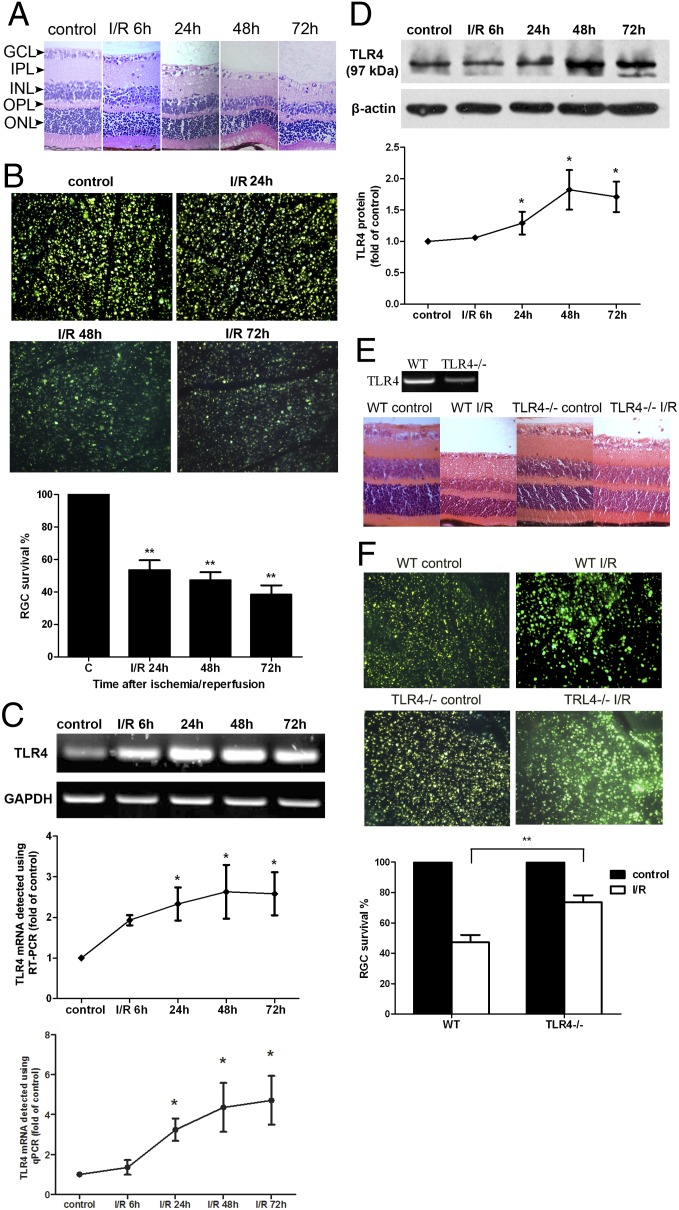
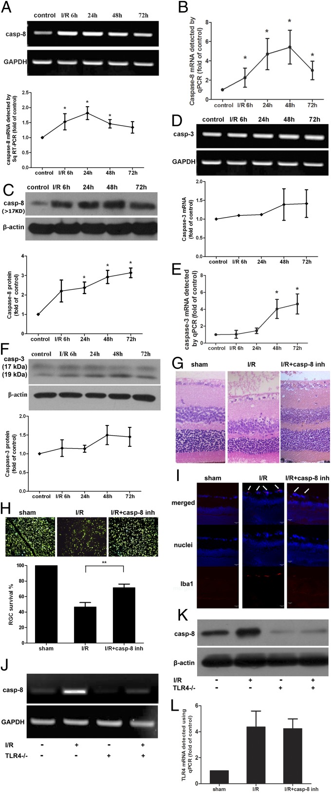
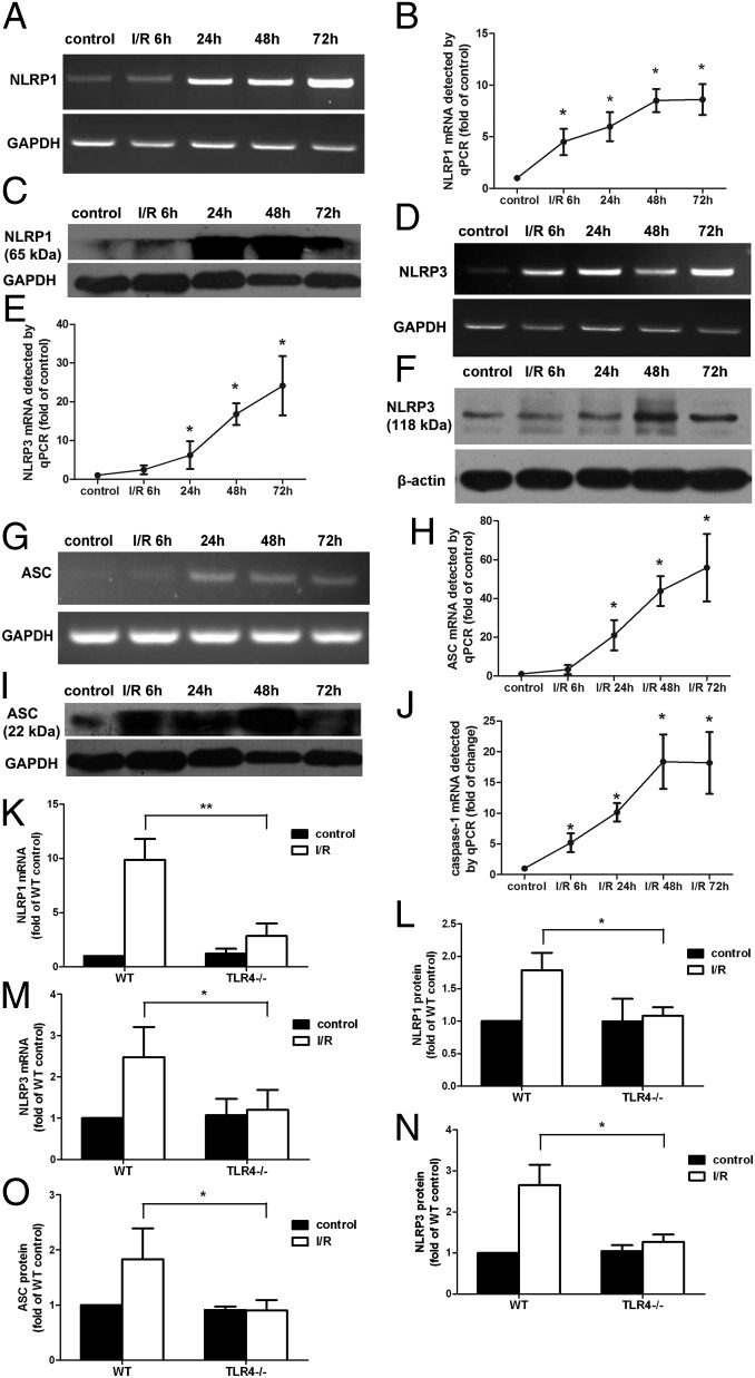
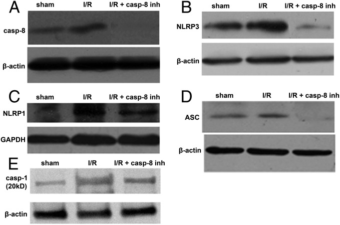
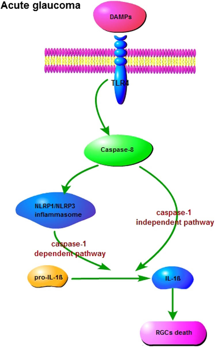
Acute glaucoma is a sight-threatening condition characterized by a sudden and substantial rise in intraocular pressure (IOP) and consequent retinal ganglion cell (RGC) death. Angle closure glaucoma, a common cause of glaucoma in Asia that affects tens of millions of people worldwide, often presents acutely with loss of vision, pain, and high IOP. Even when medical and surgical treatment is available, acute angle closure glaucoma can cause permanent and irreversible loss of vision. Toll-like receptor 4 (TLR4) signaling has been previously implicated in the pathogenesis of IOP-induced RGC death, although the underlying mechanisms are largely unknown. In the present study, we used an acute IOP elevation/glaucoma model to investigate the underlying mechanism of RGC death. We found that TLR4 leads to increased caspase-8 expression; this elevation increases IL-1β expression and RGC death via a caspase-1-dependent pathway involving Nod-like receptor family, pyrin domain containing 1 (NLRP1)/NLRP3 inflammasomes and a caspase-1-independent pathway. We show that inhibition of caspase-8 activation significantly attenuates RGC death by down-regulating the activation of NLRP1 and NLRP3, thus demonstrating the pivotal role of caspase-8 in the TLR4-mediated activation of inflammasomes. These findings demonstrate collectively a critical role of caspase-8 in transducing TLR4-mediated IL-1β production and RGC death and highlight signal transduction in a caspase-1-dependent NLRP1/NLRP3 inflammasome pathway and a caspase-1-independent pathway in acute glaucoma. These results provide new insight into the pathogenesis of glaucoma and point to a treatment strategy.

Keywords: cell apoptosis; retinal ischemia/reperfusion injury.


Conflict of interest statement

The authors declare no conflict of interest.

Figures

Fig. 1.
Fig. 1.
TLR4 signaling was involved in the pathogenesis of IOP-induced RGC death. (A) H&E staining of retinal cross-sections showed degeneration of RGCs and a decrease in the mean thickness of retinal tissue of ischemia/reperfusion (I/R) mice at different time points after reperfusion. GCL, ganglion cell layer; INL, inner nuclear layer; IPL, inner plexiform layer; ONL, outer nuclear layer; OPL, outer plexiform layer (n = 6). (Scale bar: 10 μm.) (B) FG labeling showed that the number of surviving cells in the RGC layer decreased significantly after IOP-induced injury compared with controls (n = 12). (C) sqRT-PCR and qPCR analysis of TLR4 gene expression in ischemic retina at different time points after reperfusion. Levels of TLR4 mRNA were elevated in ischemic retina compared with controls (n = 8). (D) Western blot analysis showed that TLR4 protein levels were also up-regulated in ischemic retina after reperfusion (n = 12). (E and F) Knockdown of TLR4 reduced retinal ischemic damage and RGC death. A sham procedure was performed in the contralateral eyes, which served as controls (n = 8 each for WT and TLR4−/− mice). Representative images are shown. Data are presented as mean ± SD of the fold increase compared with controls. *P < 0.05, **P < 0.001.
Fig. 2.
Fig. 2.
Increased production of caspase-8, induced by TLR4, contributed to the pathogenesis of IOP-induced retinal damage. Caspase-8 gene (A and B) (n = 10) and protein expression (C) (n = 15) were significantly up-regulated in ischemic retina compared with controls. Caspase-3 mRNA levels (D and E) (n = 10) and protein levels (F) (n = 15) were mildly increased in ischemic retina until 48 h after reperfusion. Inhibition of caspase-8 via Z-IETD-fmk injection into the vitreous cavity significantly attenuated retinal ischemic damage (G) (n = 9), RGC death (H) (n = 9), and microglia activation (I) (n = 9). By knocking out the TLR4 gene using TLR4−/− mice, the expression of caspase-8 was reduced following IOP-induced injury (J and K) (n = 6) compared with WT mice (n = 6). Inhibition of caspase-8 had no effect on the expression of TLR4 in ischemic retina (L) (n = 6). Representative images taken 48 h after I/R injury are shown. Data are presented as mean ± SD; *P < 0.05, **P < 0.001.
Fig. 3.
Fig. 3.
The production of NLRP1 and NLRP3 inflammasomes was increased in ischemic retina, mediated by TLR4 signaling. NLRP1 and NLRP3 gene expression (A, B, D, and E) (n = 10, both) and protein expression (C and F) (n = 15, both) in ischemic retina were significantly up-regulated compared with controls. ASC mRNA levels detected using sqRT-PCR (G) and qPCR (H) (n = 6, both) and protein expression (I) (n = 15) were also up-regulated in IOP-induced retinal injury. (J) The expression of caspase-1 was correspondingly elevated in ischemic retinal tissue (n = 6). Knockdown of TLR4 reduced the production of NLRP1 and NLRP3 inflammasomes and ASC (KO) (n = 12, all). Representative images are shown. Data are presented as mean ± SD; *P < 0.05, **P < 0.001.
Fig. 4.
Fig. 4.
Caspase-8 signaling regulated the activation of NLRP1 and NLRP3 inflammasomes in IOP-induced retinal damage. The intravitreous injection of the caspase-8 inhibitor Z-IETD-fmk (20 μM) (A) significantly attenuated the activation of NLRP3 (B), NLRP1 (C), ASC (D), and caspase-1 (E) (n = 9, all) following retinal ischemic injury. Representative images are shown.
Fig. 5.
Fig. 5.
Caspase-8 signaling was required for processing of IL-1β only partially through caspase-1–dependent NLRP1 and NLRP3 inflammasome activation. Gene expression (A) (n = 10) and protein expression of IL-1β (B) (n = 15) were elevated in ischemic retina at different time points after reperfusion. (C) The intravitreous injection of caspase-8 inhibitor, Z-IETD-fmk (20 μM), completely inhibited the processing of IL-1β (n = 9). (D) The intravitreous injection of caspase-1 inhibitor, Z-YVAD-fmk (20 μM), partially reduced the processing of IL-1β (n = 9). (E and F) Inhibition of caspase-1 attenuated the damage and RGC death from retinal ischemia (n = 12). Representative images are shown. Data are presented as mean ± SD; *P < 0.05, **P < 0.001.
Fig. 6.
Fig. 6.
Diagram illustrating the proposed pathway by which TLR4/caspase-8/inflammasome up-regulation induces the processing of IL-1β and promotes the inflammatory response in acute glaucoma.

References

    1. Weinreb RN, Aung T, Medeiros FA. The pathophysiology and treatment of glaucoma: A review. JAMA. 2014;311(18):1901–1911. - PMC - PubMed
    1. Wang N, Wu H, Fan Z. Primary angle closure glaucoma in Chinese and Western populations. Chin Med J (Engl) 2002;115(11):1706–1715. - PubMed
    1. Ang LP, Ang LP. Current understanding of the treatment and outcome of acute primary angle-closure glaucoma: An Asian perspective. Ann Acad Med Singapore. 2008;37(3):210–215. - PubMed
    1. Quek DT, et al. Blindness and long-term progression of visual field defects in chinese patients with primary angle-closure glaucoma. Am J Ophthalmol. 2011;152(3):463–469. - PubMed
    1. Ousman SS, Kubes P. Immune surveillance in the central nervous system. Nat Neurosci. 2012;15(8):1096–1101. - PMC - PubMed

Publication types

MeSH terms

Substances