. 2014 Jul 29;111(30):11061-6.
doi: 10.1073/pnas.1322362111. Epub 2014 Jul 14.

Arhgap36-dependent activation of Gli transcription factors

Affiliations

Arhgap36-dependent activation of Gli transcription factors

Paul G Rack et al. Proc Natl Acad Sci U S A. .

Abstract

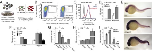
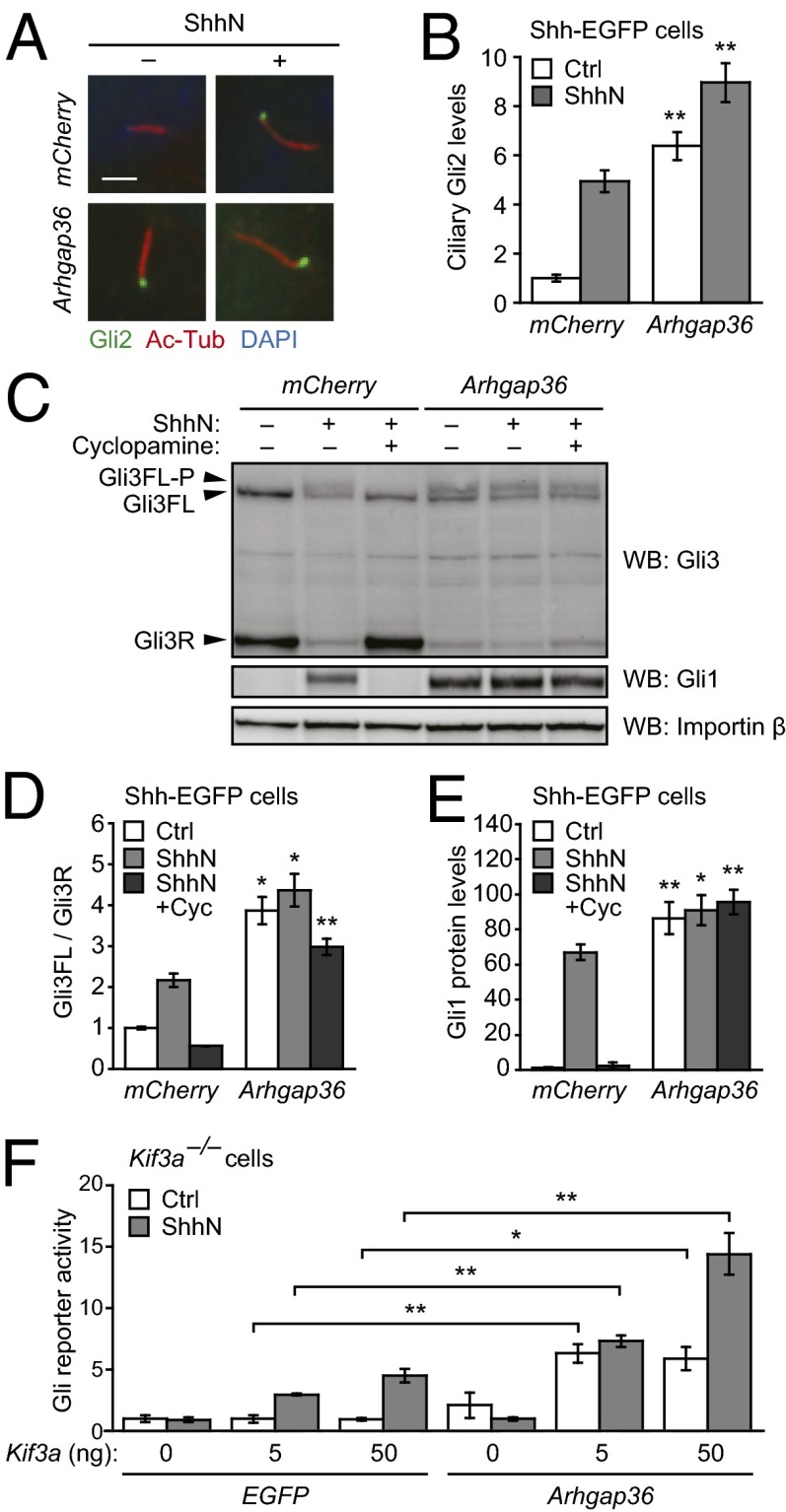
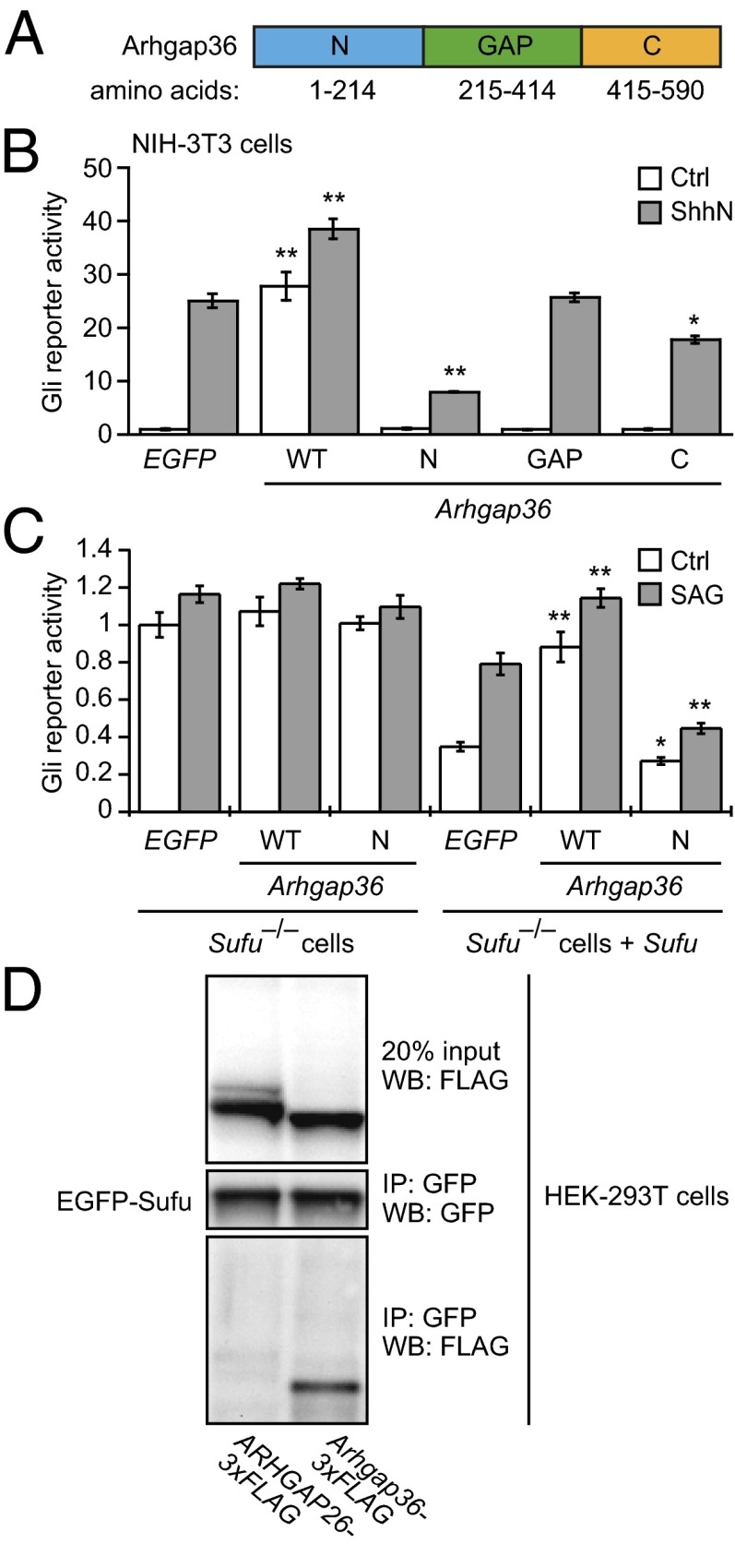
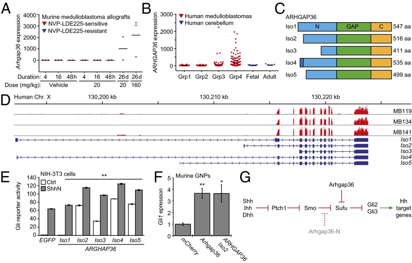
Hedgehog (Hh) pathway activation and Gli-dependent transcription play critical roles in embryonic patterning, tissue homeostasis, and tumorigenesis. By conducting a genome-scale cDNA overexpression screen, we have identified the Rho GAP family member Arhgap36 as a positive regulator of the Hh pathway in vitro and in vivo. Arhgap36 acts in a Smoothened (Smo)-independent manner to inhibit Gli repressor formation and to promote the activation of full-length Gli proteins. Arhgap36 concurrently induces the accumulation of Gli proteins in the primary cilium, and its ability to induce Gli-dependent transcription requires kinesin family member 3a and intraflagellar transport protein 88, proteins that are essential for ciliogenesis. Arhgap36 also functionally and biochemically interacts with Suppressor of Fused. Transcriptional profiling further reveals that Arhgap36 is overexpressed in murine medulloblastomas that acquire resistance to chemical Smo inhibitors and that ARHGAP36 isoforms capable of Gli activation are up-regulated in a subset of human medulloblastomas. Our findings reveal a new mechanism of Gli transcription factor activation and implicate ARHGAP36 dysregulation in the onset and/or progression of GLI-dependent cancers.


Conflict of interest statement

The authors declare no conflict of interest.

Figures

Fig. 1.
Fig. 1.
Identification of Arhgap36 as a Hh pathway activator. (A) Schematic representation of the cDNA overexpression screen. (B) FACS scatter plots revealing a retroviral pool that induces Hh ligand-independent pathway activation. Retroviral transduction of mCherry alone served as a negative control, and the percentage of total cells within the sorting gate (dashed box) for each condition is indicated. Deconvolution of this sublibrary identified ARHGAP36 as the active component. (C) FACS histogram plots demonstrating the response of Shh-EGFP cells to ShhN or exogenous Arhgap36. (D) Alkaline phosphatase (AP) levels in C3H10T1/2 cells transduced with mCherry or Arhgap36 and then cultured in the absence or presence of ShhN for 48 h. Data are the average ± SEM, n = 8. (E) Expression levels of ptch2 in uninjected zebrafish embryos (Top) or those injected with Arhgap36 (Middle) or dnPKA (Bottom) mRNA. Lateral views of 24-hpf embryos are shown. (Scale bar: 200 μm.) (F) Hh pathway activities in NIH-3T3 cells cotransfected with Gli-dependent firefly luciferase and SV40-driven Renilla luciferase reporters and either EGFP or Arhgap36. The cells were then treated with ShhN, 200 nM SAG, or 5 µM cyclopamine for 30 h. Data are the average ± SEM, n = 3. (G) Hh pathway activities in Smo−/− MEFs cotransfected with the luciferase reporters and designated cDNAs. (H) Hh pathway activities in Gli2−/− MEFs cotransfected with the luciferase reporters and designated cDNAs for 48 h. (I) Hh pathway activities in Gli3−/− MEFs cotransfected with the luciferase reporters and designated cDNAs and then cultured in the absence of presence of ShhN for 30 h. Data for GI are the average ± SEM, n = 4. Statistical analyses: Single and double asterisks indicate P < 0.05 and P < 0.01, respectively (D, Arhgap36 vs. mCherry for each cell culture condition; F, Arhgap36 vs. EGFP for each cell culture condition; GI, each designated gene versus EGFP).
Fig. 2.
Fig. 2.
Effects of Arhgap36 on Gli trafficking and processing. (A) Ciliary Gli2 levels in Shh-EGFP cells retrovirally transduced with either mCherry or Arhgap36 and then cultured in the absence or presence of ShhN for 1 h. Representative immunofluorescence micrographs are shown with staining for Gli2, acetylated tubulin (primary cilia), and DAPI (nuclei). (Scale bar: 2 µm.) (B) Quantification of Gli2 immunofluorescence within primary cilia. Data are the average ± SEM, n ≥ 50 cells. (C) Gli3 forms in Shh-EGFP cells transduced with either mCherry or Arhgap36 and concurrently treated with ShhN-free medium, ShhN, or ShhN plus 5 µM cyclopamine for 24 h. A representative blot is shown with protein bands corresponding to Gli3R, Gli3FL, and the phosphorylated form of Gli3FL labeled. Gli1 levels indicate Hh pathway state, and importin β levels were used as gel-loading controls. (D and E) Quantification of Gli3FL/Gli3R ratios and Gli1 levels for each experimental condition. Data are the average ± SEM, n = 3 independent blots. (F) Hh pathway activities in Kif3a−/− MEFs cotransfected with Gli-dependent firefly luciferase and SV40-driven Renilla luciferase reporters, either EGFP or Arhgap36, and varying amounts of exogenous Kif3a. The cells were then cultured in absence or presence of ShhN for 30 h. Data are the average ± SEM, n = 4. Statistical analyses: Single and double asterisks indicate P < 0.05 and P < 0.01, respectively (AE, Arhgap36 versus mCherry for each cell culture condition; F, Arhgap36 versus EGFP for each Kif3a transfection and cell culture condition).
Fig. 3.
Fig. 3.
Arhgap36 domain analysis and interactions with Sufu. (A) Schematic representation of Arhgap36 with the N-terminal, Rho GAP, and C-terminal domains labeled. (B) Hh pathway activities in NIH-3T3 cells cotransfected with Gli-dependent firefly luciferase and SV40-driven Renilla luciferase reporters and EGFP, wild-type Arhgap36, or the indicated Arhgap36 domains. Data are the average ± SEM, n = 3. (C) Hh pathway activities in Sufu−/− MEFs cotransfected with the luciferase reporters and either EGFP or the designated cDNAs. Sufu and Sufu/SAG were used to verify Sufu-dependent Arhgap36 function and the Hh pathway-competence of the cells. Data are the average ± SEM, n = 4. (D) Immunoprecipitation studies of HEK-293T cells cotransfected with EGFP-Sufu and either ARHGAP26-3xFLAG or Arhgap36-3xFLAG. Statistical analyses: Single and double asterisks indicate P < 0.05 and P < 0.01, respectively (Arhgap36 or its individual domains versus EGFP for each cell culture condition).
Fig. 4.
Fig. 4.
ARHGAP36 up-regulation in medulloblastoma and a model for Arhgap36-dependent Hh pathway regulation. (A) Arhgap36 transcript levels in NVP-LDE225–sensitive and resistant medulloblastoma allografts derived from Ptch+/−;p53−/− mice (data from ref. 29). (B) ARHGAP36 transcript levels in human medulloblastomas (groups 1–4) and normal cerebellar tissue, as determined by microarray analysis. (C) Schematic representation of the five predicted ARHGAP36 isoforms with the N-terminal, Rho GAP, and C-terminal domains labeled. (D) Alignments of the RNA-seq read coverage for three medulloblastoma samples, the ARHGAP36 genomic locus (Genome Reference Consortium GRCh37/hg19), and exon-intron structures for the predicted ARHGAP36 isoforms (UniprotKB/Swiss-Prot Q6ZRI8KB designations). Reads for all predicted ARHGAP36 exons except for the first were observed, indicating that the tumors were only capable of generating isoforms 2, 3, and 5. (E) Hh pathway activities in NIH-3T3 cells cotransfected with Gli-dependent firefly luciferase and SV40-driven Renilla luciferase reporters and either EGFP or individual ARHGAP36 isoforms with C-terminal V5 tags. The cells were then cultured in the absence or presence of ShhN for 30 h. Data are the average ± SEM, n = 3, and the double asterisks indicate P < 0.01 (ARHGAP36 isoforms versus EGFP for each cell culture condition). (F) Hh pathway activities in murine cerebellar GNPs retrovirally transduced with mCherry, Arhgap36, or ARHGAP36 (isoform 2) and treated with 3 µM cyclopamine for 48 h, as measured by endogenous Gli1 expression. Data are the average ± SEM, n = 6, and the double asterisks indicate P < 0.01 (ARHGAP36 isoforms versus EGFP for each cell culture condition). (G) Schematic representation of noncanonical Hh pathway activation by Arhgap36 and the inhibition of canonical Hh signaling by Arhgap36-N. Statistical analyses: Single and double asterisks indicate P < 0.05 and P < 0.01, respectively (E, ARHGAP36 isoforms versus EGFP for each cell culture condition; F, Arhgap36 or ARHGAP36 isoform 2 versus mCherry).

References

    1. Hui CC, Angers S. Gli proteins in development and disease. Annu Rev Cell Dev Biol. 2011;27:513–537. - PubMed
    1. Zacharias WJ, et al. Hedgehog signaling controls homeostasis of adult intestinal smooth muscle. Dev Biol. 2011;355(1):152–162. - PMC - PubMed
    1. Teglund S, Toftgård R. Hedgehog beyond medulloblastoma and basal cell carcinoma. Biochim Biophys Acta. 2010;1805(2):181–208. - PubMed
    1. Tukachinsky H, Lopez LV, Salic A. A mechanism for vertebrate Hedgehog signaling: Recruitment to cilia and dissociation of SuFu-Gli protein complexes. J Cell Biol. 2010;191(2):415–428. - PMC - PubMed
    1. Humke EW, Dorn KV, Milenkovic L, Scott MP, Rohatgi R. The output of Hedgehog signaling is controlled by the dynamic association between Suppressor of Fused and the Gli proteins. Genes Dev. 2010;24(7):670–682. - PMC - PubMed

Publication types

MeSH terms

Associated data