. 2014:2014:468285.
doi: 10.1155/2014/468285. Epub 2014 Jun 12.

The impact of Staphylococcus aureus-associated molecular patterns on staphylococcal superantigen-induced toxic shock syndrome and pneumonia

Affiliations

The impact of Staphylococcus aureus-associated molecular patterns on staphylococcal superantigen-induced toxic shock syndrome and pneumonia

Ashenafi Y Tilahun et al. Mediators Inflamm. 2014.

Abstract

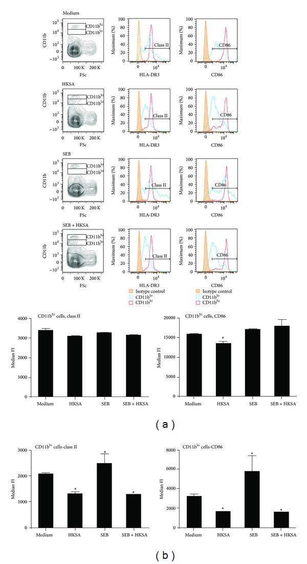
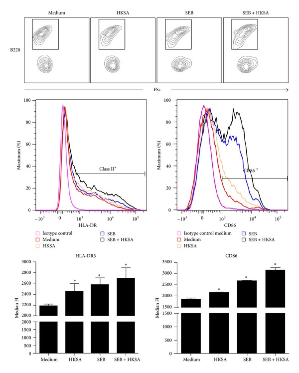
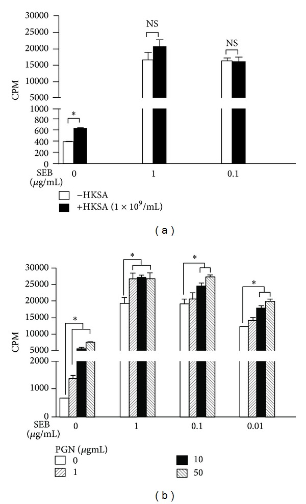
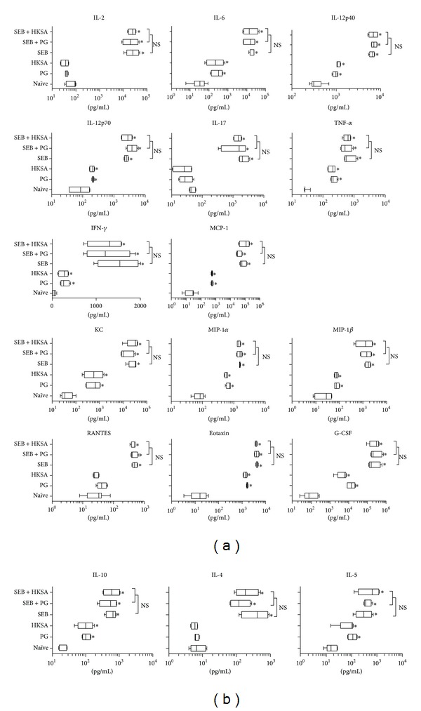
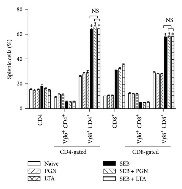
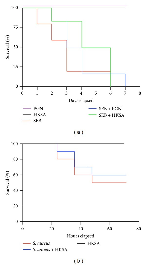
Staphylococcus aureus is capable of causing a spectrum of human illnesses. During serious S. aureus infections, the staphylococcal pathogen-associated molecular patterns (PAMPs) such as peptidoglycan, lipoteichoic acid, and lipoproteins and even intact S. aureus, are believed to act in conjunction with the staphylococcal superantigens (SSAg) to activate the innate and adaptive immune system, respectively, and cause immunopathology. However, recent studies have shown that staphylococcal PAMPs could suppress inflammation by several mechanisms and protect from staphylococcal toxic shock syndrome, a life-threatening systemic disease caused by toxigenic S. aureus. Given the contradictory pro- and anti-inflammatory roles of staphylococcal PAMPs, we examined the effects of S. aureus-derived molecular patterns on immune responses driven by SSAg in vivo using HLA-DR3 and HLA-DQ8 transgenic mice. Our study showed that neither S. aureus-derived peptidoglycans (PGN), lipoteichoic acid (LTA), nor heat-killed Staphylococcus aureus (HKSA) inhibited SSAg-induced T cell proliferation in vitro. They failed to antagonize the immunostimulatory effects of SSAg in vivo as determined by their inability to attenuate systemic cytokine/chemokine response and reduce SSAg-induced T cell expansion. These staphylococcal PAMPs also failed to protect HLA-DR3 as well as HLA-DQ8 transgenic mice from either SSAg-induced toxic shock or pneumonia induced by a SSAg-producing strain of S. aureus.


Figures

Figure 1
Figure 1
Modulation of expression of HLA-DR3 and CD86 on CD11b+ cells by HKSA. Splenic mononuclear cells from HLA-DR3 transgenic mice were cultured with medium, HKSA (108 bacteria/mL), SEB (1 μg/mL), or SEB + HKSA. 24 hours later, the cells were harvested and washed and expression of HLA-DR3 and CD86 on CD11b+ cells was analyzed by flow cytometry. Mononuclear cells were first gated based on forward and side scatter profiles. Other analyses were performed on cells within this gate. Representative histogram profiles and bar charts depicting median fluorescent intensity (MFI) are given. Each bar represents mean ± SE from 2 different experiments, each performed in triplicate. *P < 0.05 compared to cells cultured with medium.
Figure 2
Figure 2
Modulation of expression of HLA-DR3 and CD86 on B220+ cells by HKSA. Splenic mononuclear cells from HLA-DR3 transgenic mice were cultured with medium, HKSA (108 bacteria/mL), SEB (1 μg/mL), or SEB + HKSA. 24 hours later, the cells were harvested and washed and expression of HLA-DR3 and CD86 on B220+ cells was analyzed by flow cytometry. Mononuclear cells were first gated based on forward and side scatter profiles. Other analyses were performed on cells within this gate. Representative histogram profiles and bar charts depicting median fluorescent intensity (MFI) are given. Each bar represents mean ± SE from 2 different experiments, each performed in triplicate. *P < 0.05 compared to cells cultured with medium.
Figure 3
Figure 3
Effect of S. aureus-derived PAMPs on SEB-induced T cell proliferation in vitro. Splenic mononuclear cells from HLA-DR3 transgenic mice were cultured with indicated concentrations of SEB in the presence or absence of HKSA, 108 bacteria/mL (a), or PGN (b). T cell proliferation was determined by thymidine incorporation. *P < 0.05 compared to cells cultured without HKSA or PGN within that group. Each bar represents mean ± SE from triplicate wells. Representative data from one out of 3 similar experiments are given.
Figure 4
Figure 4
Effect of S. aureus-derived PAMPs on SEB-induced systemic inflammatory response syndrome in vivo. HLA-DR3 transgenic mice were challenged with staphylococcal enterotoxin B (SEB) alone (50 μg), staphylococcal peptidoglycan (PGN) alone (50 μg), or heat-killed Staphylococcus aureus (HKSA) alone (108 bacteria), SEB with PGN or SEB with HKSA. Animals bled 4 hours after injection and serum cytokines were quantified using Bioplex assays (Bio-Rad). Mean ± SE from 4–6 mice in each group. *P < 0.05 compared to naïve mice.
Figure 5
Figure 5
Effect of S. aureus-derived PAMPs on SEB-induced T cell expansion in vivo. HLA-DR3 transgenic mice were challenged with staphylococcal enterotoxin B (SEB) alone (10 μg), staphylococcal peptidoglycan (PGN) alone (50 μg), or lipoteichoic acid (LTA, 50 μg), SEB with PGN or SEB with LTA. Animals were killed 3 days later and distribution of T cells expressing TCR Vβ families were determined by flow cytometry. Mean ± SE from 5–8 mice in each group. *P < 0.05 compared to naïve mice. p = NS between SEB versus SEB + PGN, SEB versus SEB + LTA, and SEB + LTA versus SEB + PGN.
Figure 6
Figure 6
Effect of S. aureus-derived PAMPs on SEB-induced TSS and S. aureus-induced pneumonia. (a) HLA-DR3 transgenic mice were challenged with staphylococcal enterotoxin B (SEB) (50 μg), staphylococcal peptidoglycan (PGN) (50 μg), or heat-killed Staphylococcus aureus (HKSA) (108 bacteria), SEB with PGN or SEB with HKSA. Animals were monitored every 6 hours (4–8 mice per group). (b) HLA-DR3 transgenic mice were challenged intratracheally with a toxigenic strain of S. aureus capable of producing the SSAg, SEA, and SEB (1.3–2.5 × 10⁸ cfu/mouse). Immediately following bacterial inoculation, mice were left untreated or injected with HKSA intraperitoneally (108 bacteria/mouse). Animals were monitored closely for symptoms. All moribund animals were removed from the study (6–8 mice per group).

References

    1. Kluytmans J, van Belkum A, Verbrugh H. Nasal carriage of Staphylococcus aureus: epidemiology, underlying mechanisms, and associated risks. Clinical Microbiology Reviews. 1997;10:505–520. - PMC - PubMed
    1. Klevens RM, Morrison MA, Nadle J, et al. Invasive methicillin-resistant Staphylococcus aureus infections in the United States. The Journal of the American Medical Association. 2007;298(15):1763–1771. - PubMed
    1. Otto M. Understanding the epidemic of community-associated MRSA and finding a cure: are we asking the right questions? Expert Review of Anti-Infective Therapy. 2009;7(2):141–143. - PMC - PubMed
    1. Boucher HW, Corey GR. Epidemiology of methicillin-resistant Staphylococcus aureus . Clinical Infectious Diseases. 2008;46(supplement 5):S344–S349. - PubMed
    1. Fraser JD, Proft T. The bacterial superantigen and superantigen-like proteins. Immunological Reviews. 2008;225(1):226–243. - PubMed

Publication types

MeSH terms