. 2014 Apr;4(2):143-55.
doi: 10.4103/2229-5151.134182.

Thoracostomy tubes: A comprehensive review of complications and related topics

Affiliations

Thoracostomy tubes: A comprehensive review of complications and related topics

Michael Kwiatt et al. Int J Crit Illn Inj Sci. 2014 Apr.

Abstract

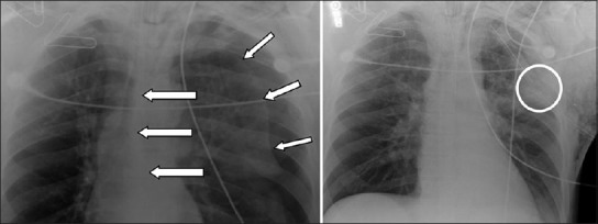
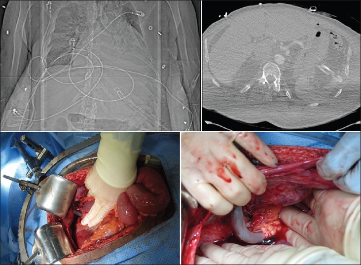
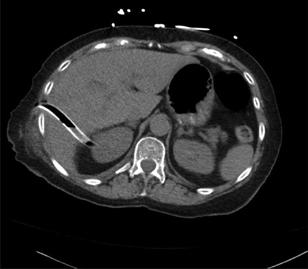
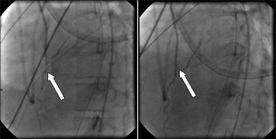
Tube thoracostomy (TT) placement belongs among the most commonly performed procedures. Despite many benefits of TT drainage, potential for significant morbidity and mortality exists. Abdominal or thoracic injury, fistula formation and vascular trauma are among the most serious, but more common complications such as recurrent pneumothorax, insertion site infection and nonfunctioning or malpositioned TT also represent a significant source of morbidity and treatment cost. Awareness of potential complications and familiarity with associated preventive, diagnostic and treatment strategies are fundamental to satisfactory patient outcomes. This review focuses on chest tube complications and related topics, with emphasis on prevention and problem-oriented approaches to diagnosis and treatment. The authors hope that this manuscript will serve as a valuable foundation for those who wish to become adept at the management of chest tubes.

Keywords: Chest tube; complications; diagnosis; prevention; review; thoracostomy tube; treatment.


Conflict of interest statement

Conflict of Interest: None declared.

Figures

Figure 1
Figure 1
Tension pneumothorax (short arrows, left) with contralateral mediastinal shift (long arrows, left). The pneumothorax was managed with emergently placed tube thoracostomy (right). Note the position of the most peripheral intake portal (circled, right). This “sentinel hole” should be located inside the thoracic cavity, and the distance markers on the chest tube indicate the distance between the “sentinel hole” and the numbered marker located at the skin level. The distance between the “sentinel hole” and the skin varies based on various factors (i.e., the position of the TT entry site on the chest wall, patient body habitus, obesity, etc)
Figure 2
Figure 2
Sub-diaphragmatic tube thoracostomy (TT) mis-placement. Plain radiograph showing the TT in the left upper quadrant (left upper image). Computed tomography confirms intra-abdominal location of the chest tube, along with free air (right upper). The patient underwent laparotomy due to the combination of both radiographic (free air) and clinical (abdominal pain) findings. Intraoperative photographs show the TT causing a superficial injury to the left hepatic lobe (left lower) and then proceeding deeper in to the left upper quadrant (peri-splenic) wrapped by omental tissue (right lower)
Figure 3
Figure 3
An example of hepatic injury secondary to thoracostomy tube. This elderly patient underwent a thoracoscopic-assisted right lower lobectomy and developed a symptomatic right-sided loculated pleural effusion several weeks later. An attempt to place right-sided chest tube at the bedside resulted in an iatrogenic injury to the hepatic parenchyma. The tube was removed in a monitored setting, with interventional radiology and surgical teams standing-by for possible hepatic parenchymal bleeding. Following tube removal, the patient remained asymptomatic and showed no signs or symptoms of bleeding
Figure 4
Figure 4
An example of postoperative chest tube compression of a coronary bypass graft (CBG).”. The chest tube is seen crossing the CBG and producing pressure-related flow-limiting lesion (arrow, left); Withdrawal of the chest tube results in improved distal flow (right)
Figure 5
Figure 5
Subcutaneous chest tube placement in a patient with a large left-sided hemothorax. Immediately upon tube thoracostomy placement, large amount of sanguineous effusion was liberated. The patient was also noted to have an air leak in the suction apparatus. The initial chest radiograph (top left) was difficult to interpret due to ECG wires interfering with proper visualization, but the tip of the chest tube (arrow) was determined to be intrathoracic. Despite improved clinical picture, both the TT drainage and the air leak quickly stopped. The subsequent CT scan shows the TT to be extrathoracic (bottom images). A new chest tube was promptly inserted and the initial chest tube removed (top right).

References

    1. Miller KS, Sahn SA. Chest tubes. Indications, technique, management and complications. Chest. 1987;91:258–64. - PubMed
    1. Ball CG, Lord J, Laupland KB, Gmora S, Mulloy RH, Ng AK, et al. Chest tube complications: How well are we training our residents? Can J Surg. 2007;50:450–8. - PMC - PubMed
    1. Antunes G, Neville E, Duffy J, Ali N. BTS guidelines for the management of malignant pleural effusions. Thora×. 2003;58(Suppl 2):29–38. - PMC - PubMed
    1. Gayer G, Rozenman J, Hoffmann C, Apter S, Simansky DA, Yellin A, et al. CT diagnosis of malpositioned chest tubes. Br J Radiol. 2000;73:786–90. - PubMed
    1. Millikan JS, Moore EE, Steiner E, Aragon GE, Van Way CW., 3rd Complications of tube thoracostomy for acute trauma. Am J Surg. 1980;140:738–41. - PubMed