

Robust Radiomics feature quantification using semiautomatic volumetric segmentation
- PMID: 25025374
- PMCID: PMC4098900
- DOI: 10.1371/journal.pone.0102107
Robust Radiomics feature quantification using semiautomatic volumetric segmentation
Abstract
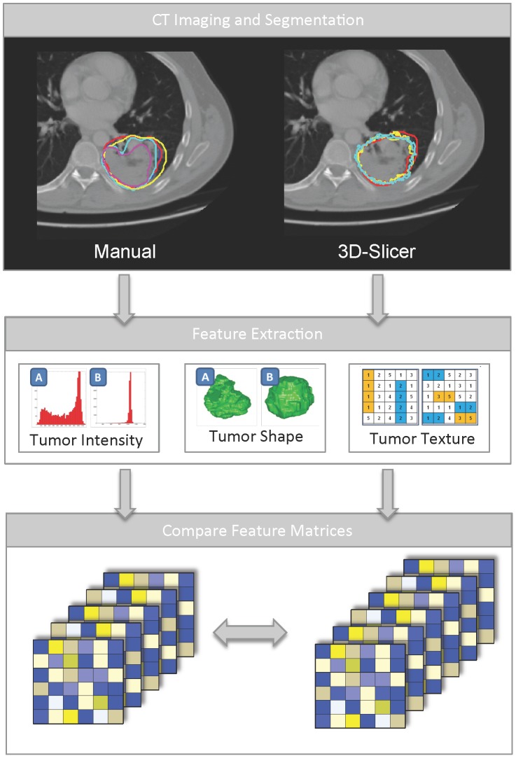
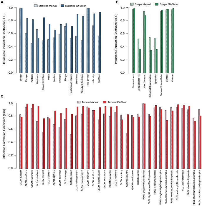
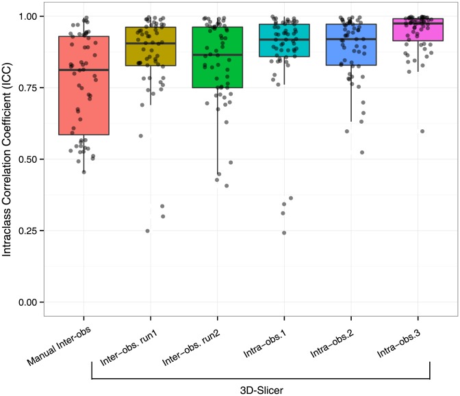
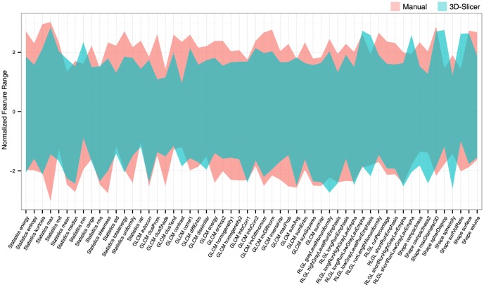
Due to advances in the acquisition and analysis of medical imaging, it is currently possible to quantify the tumor phenotype. The emerging field of Radiomics addresses this issue by converting medical images into minable data by extracting a large number of quantitative imaging features. One of the main challenges of Radiomics is tumor segmentation. Where manual delineation is time consuming and prone to inter-observer variability, it has been shown that semi-automated approaches are fast and reduce inter-observer variability. In this study, a semiautomatic region growing volumetric segmentation algorithm, implemented in the free and publicly available 3D-Slicer platform, was investigated in terms of its robustness for quantitative imaging feature extraction. Fifty-six 3D-radiomic features, quantifying phenotypic differences based on tumor intensity, shape and texture, were extracted from the computed tomography images of twenty lung cancer patients. These radiomic features were derived from the 3D-tumor volumes defined by three independent observers twice using 3D-Slicer, and compared to manual slice-by-slice delineations of five independent physicians in terms of intra-class correlation coefficient (ICC) and feature range. Radiomic features extracted from 3D-Slicer segmentations had significantly higher reproducibility (ICC = 0.85±0.15, p = 0.0009) compared to the features extracted from the manual segmentations (ICC = 0.77±0.17). Furthermore, we found that features extracted from 3D-Slicer segmentations were more robust, as the range was significantly smaller across observers (p = 3.819e-07), and overlapping with the feature ranges extracted from manual contouring (boundary lower: p = 0.007, higher: p = 5.863e-06). Our results show that 3D-Slicer segmented tumor volumes provide a better alternative to the manual delineation for feature quantification, as they yield more reproducible imaging descriptors. Therefore, 3D-Slicer can be employed for quantitative image feature extraction and image data mining research in large patient cohorts.
Conflict of interest statement
Figures
References
-
- Jemal A, Bray F, Center MM, Ferlay J, Ward E, et al. (2011) Global cancer statistics. CA: A Cancer Journal for Clinicians 61: 69–90. - PubMed
-
- van Baardwijk A, Wanders S, Boersma L, Borger J, Öllers M, et al. (2010) Mature results of an individualized radiation dose prescription study based on normal tissue constraints in stages I to III non–small-cell lung cancer. Journal of Clinical Oncology 28: 1380–1386. - PubMed
-
- Vaidya M, Creach KM, Frye J, Dehdashti F, Bradley JD, et al. (2012) Combined PET/CT image characteristics for radiotherapy tumor response in lung cancer. Radiotherapy and Oncology 102: 239–245. - PubMed
Publication types
MeSH terms
Grants and funding
LinkOut - more resources
Full Text Sources
Other Literature Sources
Medical
