

Succinate dehydrogenase (SDH)-deficient renal carcinoma: a morphologically distinct entity: a clinicopathologic series of 36 tumors from 27 patients
- PMID: 25025441
- PMCID: PMC4229399
- DOI: 10.1097/PAS.0000000000000292
Succinate dehydrogenase (SDH)-deficient renal carcinoma: a morphologically distinct entity: a clinicopathologic series of 36 tumors from 27 patients
Abstract
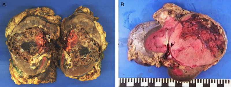
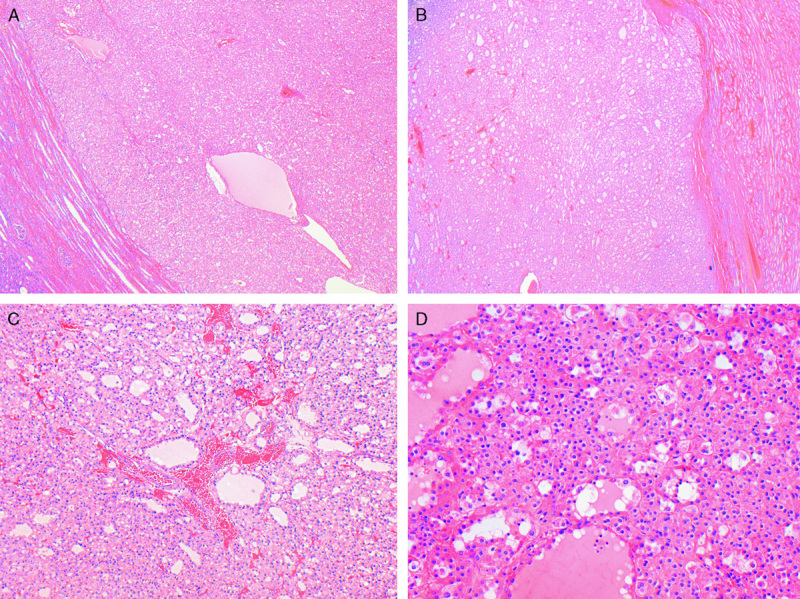
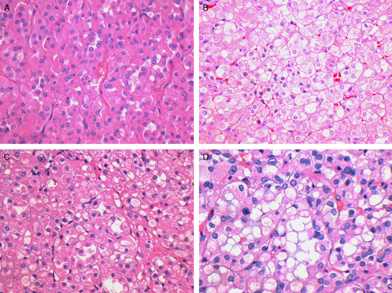
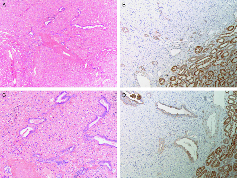
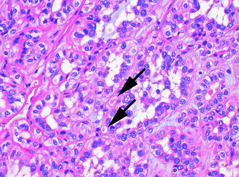
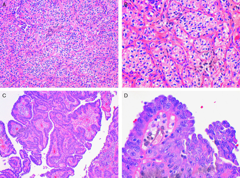
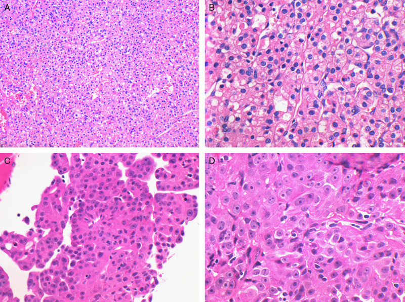
Succinate dehydrogenase (SDH)-deficient renal carcinoma has been accepted as a provisional entity in the 2013 International Society of Urological Pathology Vancouver Classification. To further define its morphologic and clinical features, we studied a multi-institutional cohort of 36 SDH-deficient renal carcinomas from 27 patients, including 21 previously unreported cases. We estimate that 0.05% to 0.2% of all renal carcinomas are SDH deficient. Mean patient age at presentation was 37 years (range, 14 to 76 y), with a slight male predominance (M:F=1.7:1). Bilateral tumors were observed in 26% of patients. Thirty-four (94%) tumors demonstrated the previously reported morphology at least focally, which included: solid or focally cystic growth, uniform cytology with eosinophilic flocculent cytoplasm, intracytoplasmic vacuolations and inclusions, and round to oval low-grade nuclei. All 17 patients who underwent genetic testing for mutation in the SDH subunits demonstrated germline mutations (16 in SDHB and 1 in SDHC). Nine of 27 (33%) patients developed metastatic disease, 2 of them after prolonged follow-up (5.5 and 30 y). Seven of 10 patients (70%) with high-grade nuclei metastasized as did all 4 patients with coagulative necrosis. Two of 17 (12%) patients with low-grade nuclei metastasized, and both had unbiopsied contralateral tumors, which may have been the origin of the metastatic disease. In conclusion, SDH-deficient renal carcinoma is a rare and unique type of renal carcinoma, exhibiting stereotypical morphologic features in the great majority of cases and showing a strong relationship with SDH germline mutation. Although this tumor may undergo dedifferentiation and metastasize, sometimes after a prolonged delay, metastatic disease is rare in the absence of high-grade nuclear atypia or coagulative necrosis.
Conflict of interest statement
Conflicts of Interest and Source of Funding: Supported in part by the Cancer Institute New South Wales and Czech Republic Government grant agency (IGA NT 12010-4). The authors have disclosed that they have no significant relationships with, or financial interest in, any commercial companies pertaining to this article.
Figures
References
-
- Gill AJ. Succinate dehydrogenase (SDH) and mitochondrial driven neoplasia. Pathology. 2012;44:285–292. - PubMed
-
- Gill AJ, Chou A, Vilain R, et al. Immunohistochemistry for SDHB divides gastrointestinal stromal tumors (GISTs) into 2 distinct types. Am J Surg Pathol. 2010;34:636–644. - PubMed
-
- Gill AJ, Pachter NS, Chou A, et al. Renal tumors associated with germline SDHB mutation show distinctive morphology. Am J Surg Pathol. 2011;35:1578–1585. - PubMed
Publication types
MeSH terms
Substances
Grants and funding
LinkOut - more resources
Full Text Sources
Other Literature Sources
Medical
