. 2015 Mar;21(3):253-62.
doi: 10.1089/ten.TEC.2014.0206. Epub 2014 Aug 19.

Autologous platelet-rich plasma: a biological supplement to enhance adipose-derived mesenchymal stem cell expansion

Affiliations

Autologous platelet-rich plasma: a biological supplement to enhance adipose-derived mesenchymal stem cell expansion

Fatemeh Atashi et al. Tissue Eng Part C Methods. 2015 Mar.

Abstract

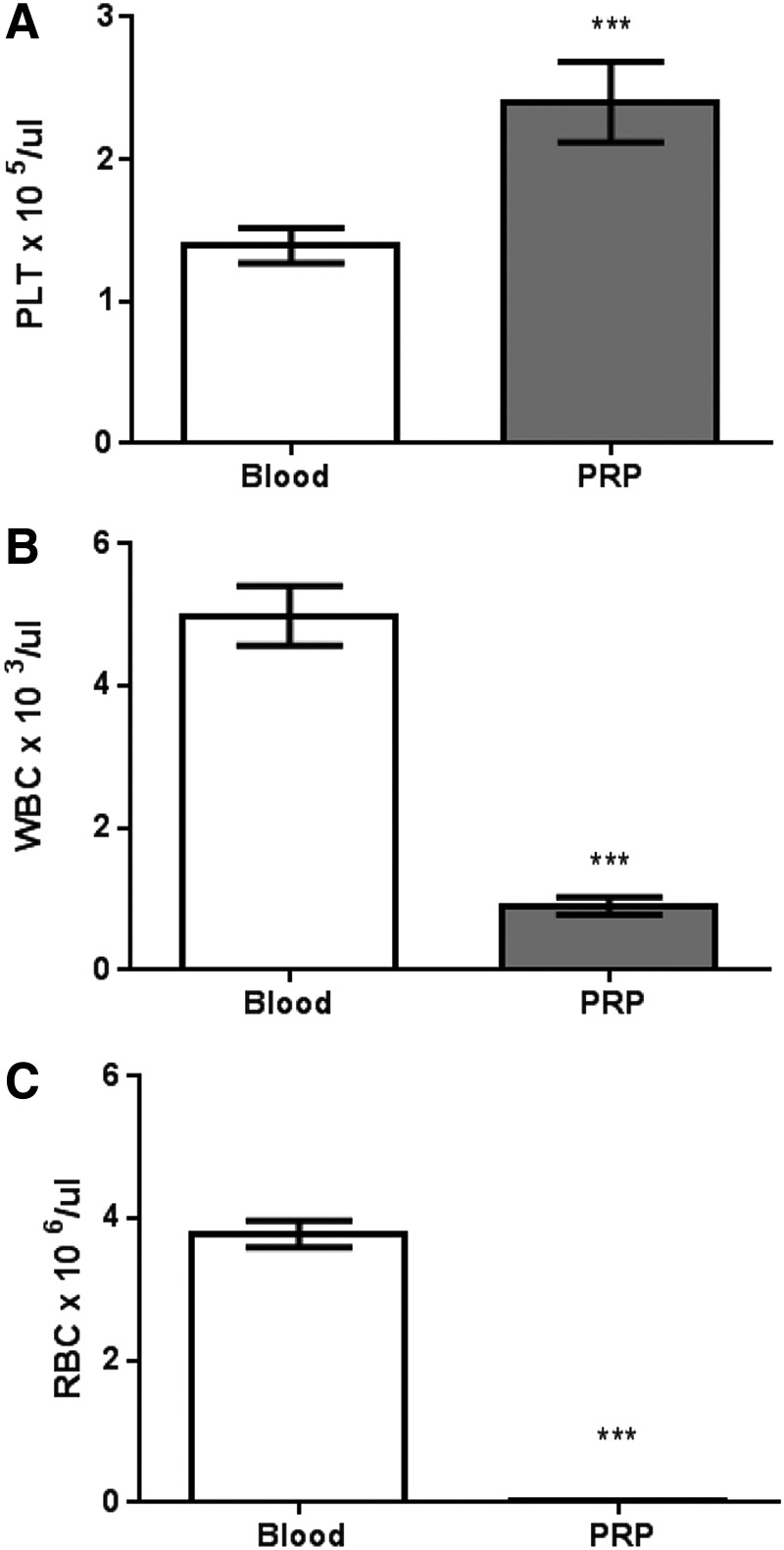
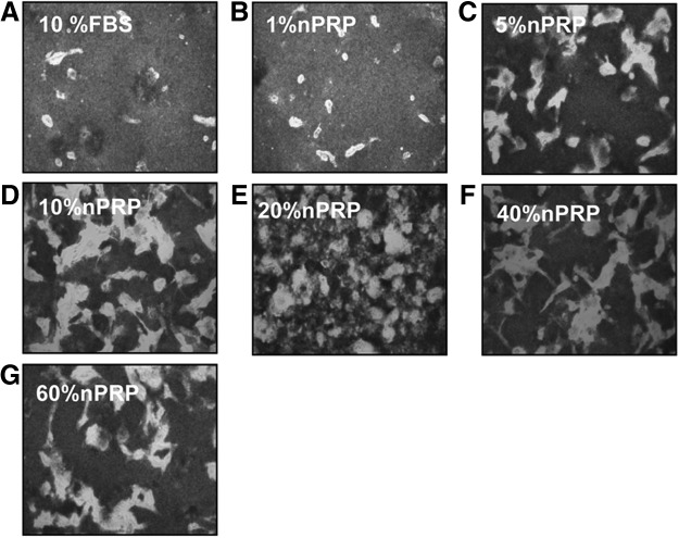
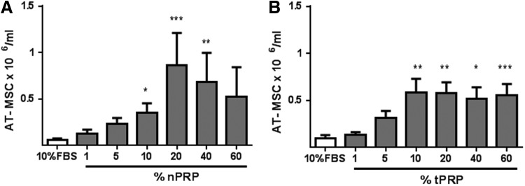
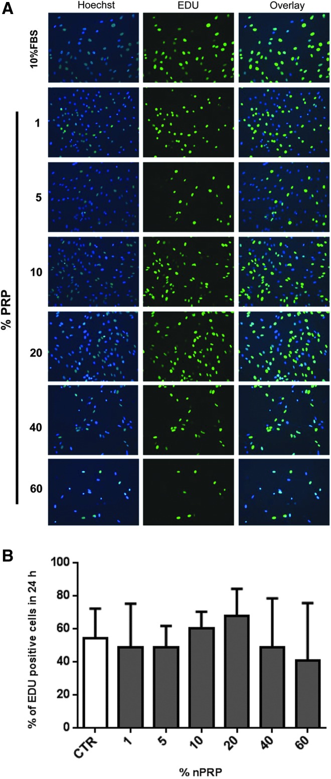
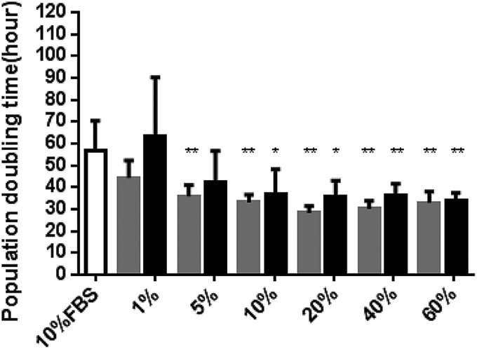
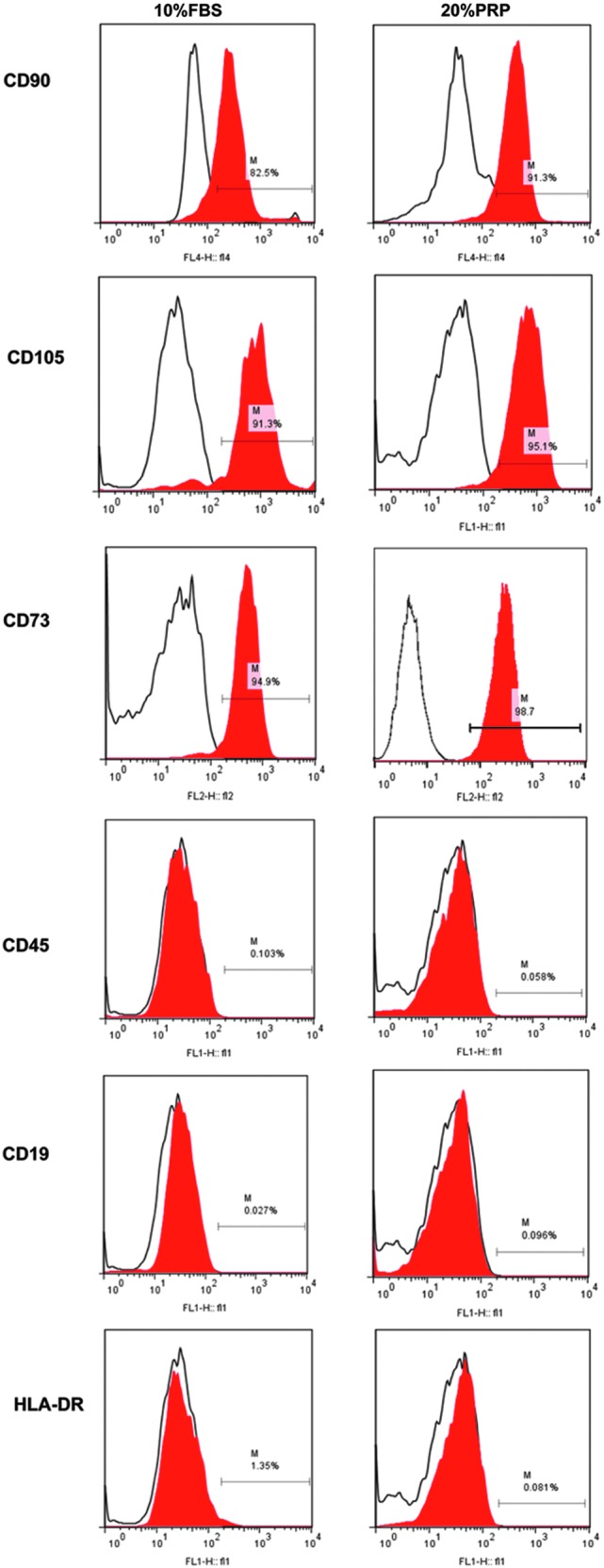
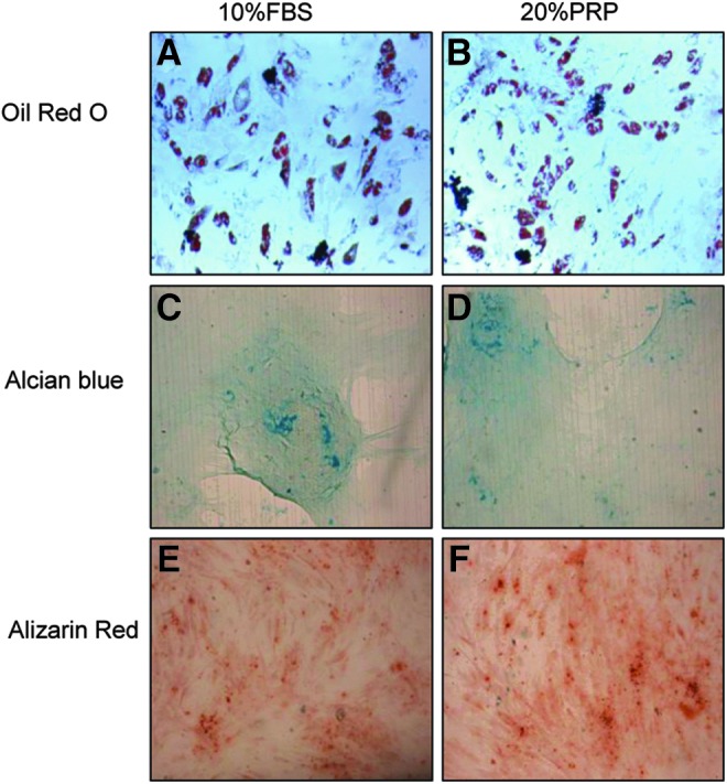
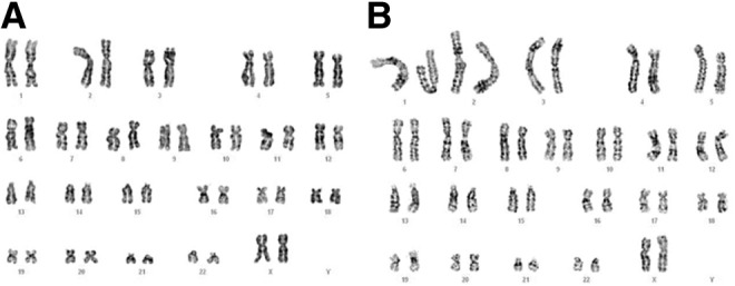
Currently the use of non-autologous cell culture media (e.g., animal-derived or allogeneic serum) for clinical applications of mesenchymal stem cells (MSCs) is criticized by regulatory agencies. Autologous platelet-rich plasma (PRP) is proposed as a safer alternative medium supplement for adipose-derived mesenchymal stem cells (AT-MSC) culture. To study its efficiency on cell proliferation, AT-MSCs were cultured for 10 days in media supplemented with different concentrations of autologous non-activated PRP (nPRP) or thrombin-activated PRP (tPRP) (1-60%). AT-MSC proliferation, cell phenotype, multipotency capacity, and chromosome stability were assessed and compared to AT-MSCs expanded in a classical medium supplemented with 10% of fetal bovine serum (FBS). Culture media supplemented with nPRP showed dose-dependent higher AT-MSC proliferation than did FBS or tPRP. Twenty percent nPRP was the most effective concentration to promote cell proliferation. This condition increased 13.9 times greater AT-MSC number in comparison to culture with FBS, without changing the AT-MSC phenotype, differentiation capacity, and chromosome status. We concluded that 20% autologous nPRP is a safe, efficient, and cost-effective supplement for AT-MSC expansion. It should be considered as an alternative to FBS or other nonautologous blood derivatives. It could serve as a potent substitute for the validation of future clinical protocols as it respects good manufacturing practices and regulatory agencies' standards.


Figures

<b>FIG. 1.</b>
FIG. 1.
Analysis of number of platelets (A), white blood cells (B), and red blood cells (C) in whole blood compared to purified platelet-rich plasma (PRP). n=14 patients (***p<0.001).
<b>FIG. 2.</b>
FIG. 2.
Bright-field micrographs of adipose-derived mesenchymal stem cell (AT-MSC). In the presence of 10% fetal bovine serum (FBS) (A), and 1–60% nonactivated PRP (nPRP) (B–G) after 10 days culture (P0). Magnification 20×. Pictures are representatives of one donor. High density of platelets produces a darker background.
<b>FIG. 3.</b>
FIG. 3.
Effect of different concentrations of nPRP (A, n=14) or thrombin-activated PRP (tPRP) (B, n=6) on the proliferation of AT-MSC cultured over 10 days without medium change. Ten percent FBS is used as control. *p<0.05; **p<0.01 and ***p<0.001.
<b>FIG. 4.</b>
FIG. 4.
Assessment of 24 h-proliferation efficiency of AT-MSCs by EdU staining. Different concentrations of nPRP (1–60%) were compared to 10% FBS on AT-MSCs (P1) at the end of 10 days culture period. (A) Active proliferating cells are revealed by EdU-based green fluorescence nucleus staining, compared to total cells stained by Hoechst dye (blue nucleus staining). (B) Percentage of EdU positive cells was estimated by the formula: (green bright AT-MSC nucleus/total blue AT-MSC nucleus)×100. n=4.
<b>FIG. 5.</b>
FIG. 5.
Population doubling time in AT-MSC. Cells were cultured in media supplemented with 10% FBS as control (white bar), and increasing concentrations of nPRP (gray bars) or tPRP (black bars). *p<0.05 and **p<0.01 (n=6).
<b>FIG. 6.</b>
FIG. 6.
Analysis of surface marker expression. Phenotype of AT-MSCs cultured in 10% FBS or 20% nPRP was assessed by flow cytometry at passage 0. Isotype control antibody is shown by the black empty lines whereas the expressed markers are in red. “M bars” indicate the percentage of cells expressing the surface marker.
<b>FIG. 7.</b>
FIG. 7.
Representative qualitative evaluation of AT-MSC differentiation capacity. Differentiation toward adipocyte (A, B), chondrocyte (C, D), or (E, F) osteocyte phenotype after culture in 10% FBS or 20% nPRP.
<b>FIG. 8.</b>
FIG. 8.
Representative chromosome karyotype by G-banding. (A) 10% FBS and (B) 20% nPRP cultured MSCs were compared for chromosome aberration.
<b>FIG. 9.</b>
FIG. 9.
Assessment of platelet viability by Fluorescence microscopy using calcein dye. (A–D) Fluorescence micrograph of viable, calcein-stained platelets monitored over a 10 days period in media containing 20% nPRP in contact with AT-MSC without medium change (magnification: 40×). (E) Mean viable platelet in the presence or absence of AT-MSC. Percentage of platelet viability in 20% nPRP was normalized to day 0 (n=5; *p<0.05, **p<0.01).

References

    1. Braghirolli D.I., Zamboni F., Chagastelles P.C., Moura D.J., Saffi J., Henriques J.A., et al. . Bio-electrospraying of human mesenchymal stem cells: An alternative for tissue engineering. Biomicrofluidics 7,44130, 2013 - PMC - PubMed
    1. Rahman M.M., Subramani J., Ghosh M., Denninger J.K., Takeda K., Fong G.H., et al. . CD13 promotes mesenchymal stem cell-mediated regeneration of ischemic muscle. Front Physiol 4,402, 2014 - PMC - PubMed
    1. Zomorodian E., and Baghaban Eslaminejad M.Mesenchymal stem cells as a potent cell source for bone regeneration. Stem Cells Int 2012,980353, 2012 - PMC - PubMed
    1. Van Pham P., Bui K.H., Ngo D.Q., Vu N.B., Truong N.H., Phan N.L., et al. . Activated platelet-rich plasma improves adipose-derived stem cell transplantation efficiency in injured articular cartilage. Stem Cell Res Ther 4,91, 2013 - PMC - PubMed
    1. Zhou Y., Yan Z., Zhang H., Lu W., Liu S., Huang X., et al. . Expansion and delivery of adipose-derived mesenchymal stem cells on three microcarriers for soft tissue regeneration. Tissue Eng Part A 17,2981, 2011 - PubMed

Publication types