. 2014 Jul 30;5(14):5736-49.
doi: 10.18632/oncotarget.2169.

H-Prune through GSK-3β interaction sustains canonical WNT/β-catenin signaling enhancing cancer progression in NSCLC

Affiliations

H-Prune through GSK-3β interaction sustains canonical WNT/β-catenin signaling enhancing cancer progression in NSCLC

Marianeve Carotenuto et al. Oncotarget. .

Abstract

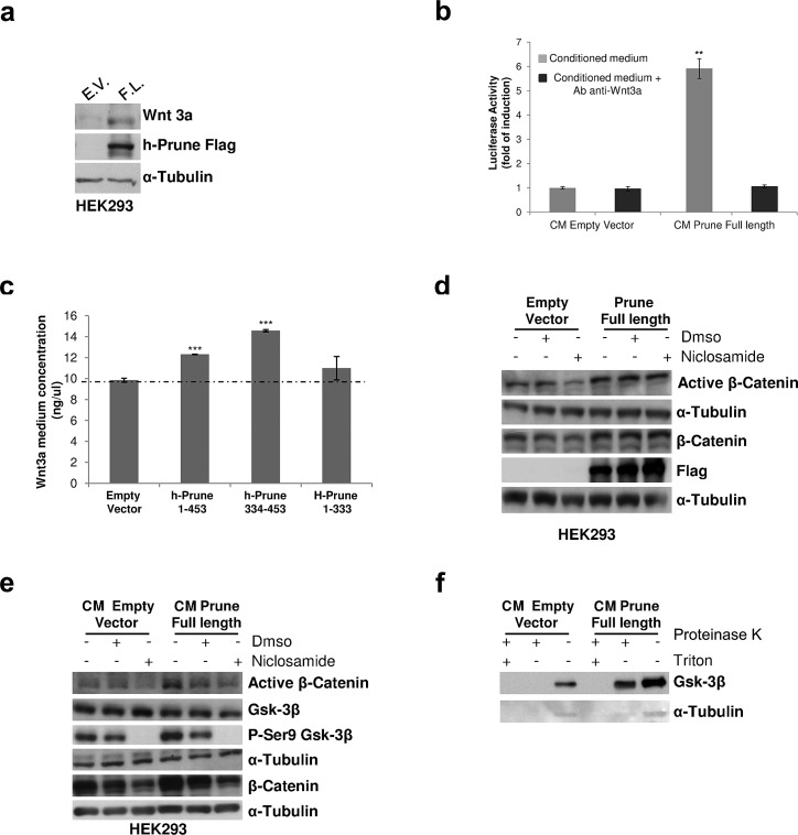
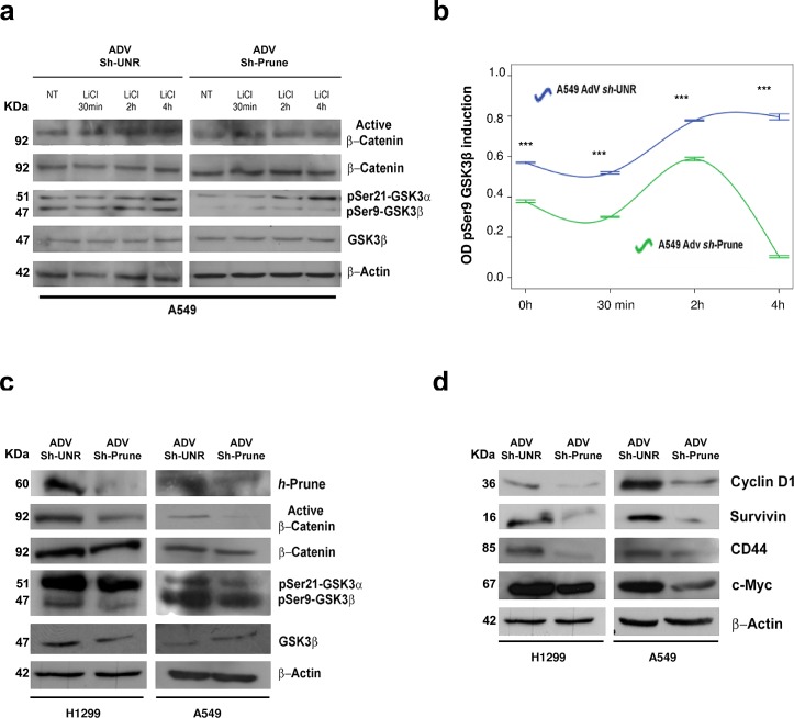
H-Prune hydrolyzes short-chain polyphosphates (PPase activity) together with an hitherto cAMP-phosphodiesterase (PDE), the latest influencing different human cancers by its overexpression. H-Prune promotes cell migration in cooperation with glycogen synthase kinase-3 (Gsk-3β). Gsk-3β is a negative regulator of canonical WNT/β-catenin signaling. Here, we investigate the role of Gsk-3β/h-Prune complex in the regulation of WNT/β-catenin signaling, demonstrating the h-Prune capability to activate WNT signaling also in a paracrine manner, through Wnt3a secretion. In vivo study demonstrates that h-Prune silencing inhibits lung metastasis formation, increasing mouse survival. We assessed h-Prune levels in peripheral blood of lung cancer patients using ELISA assay, showing that h-Prune is an early diagnostic marker for lung cancer. Our study dissects out the mechanism of action of h-Prune in tumorigenic cells and also sheds light on the identification of a new therapeutic target in non-small-cell lung cancer.


Figures

Figure 1
Figure 1. H-Prune expression enhances Wnt pathway activation
(a) Full-length h-Prune (aa 1-453) and h-Prune C-terminal region (aa 334-453) stimulation of HEK293 cells induces β-catenin-mediated transcriptional activity. The cells were transfected with the TOP/FOP flash plasmid and the Renilla luciferase plasmid overnight. The data were normalized to Renilla luciferase activity. Full-length h-Prune and h-Prune C-terminal region increased TCF transcriptional activity in HEK293 cells compared to empty vector and h-Prune N-terminal region (1-333). (b) TOP/FOP luciferase assay that shows that h-Prune induces β-catenin–mediated transcriptional activity through a GSK-3β–dependent mechanism in HEK293 cells. The cells were co-transfected with the TOP/FOP luciferase reporter, the renilla luciferase plasmid, and the control or h-Prune vector. A day after infection, the cells were treated overnight with vehicle, SB216763 (10 μM), Chiron99021 (10 μM) and IGF-1 (1 ng/ml per 5h). The data were normalized to Renilla luciferase activity and are expressed as fold-increase over vector-transfected control cells, expressed as means ±SD of three independent experiments, each carried out in triplicate. (c) Total protein prepared from HEK293 cells transfected with control vector or full-length h-Prune, which were exposed to NH4Cl (30 mM) for different times, and were subjected to Western blotting with an anti-GSK3-β antibody. α-Tubulin was used as the loading control, and an anti-Flag antibody provided the transfection control. (d) Western blots of protease protection assay in empty-vector-and h-Prune-transfected HEK293 cells. Cells were digitonin-permeabilized, and membranes were isolated and incubated without or with proteinase K in the absence or presence of Triton X-100. Gsk-3β was protected from Proteinase K in h-Prune overexpressing cells, but only in the absence of Triton X-100. (e) Representative images from immunofluorescence analysis of HEK293 cells transfected with plasmid encoding full-length h-Prune and stained for Gsk-3β and the early endosome marker EEA1 (ii), and for h-Prune and EEA1 (i). Scale bars, 10 μm. (iii, iv) Representative images from immunofluorescence analysis of HEK293 cells transfected with the plasmid encoding full-length h-Prune, and stained for Gsk-3β and the late endosome marker Rab7 (iv), and for h-Prune and Rab7 (iii). Scale bars, 20 μm.
Figure 2
Figure 2. H-Prune–conditioned medium induces Wnt signalling activation
(a) Western blotting showing that overexpression of h-Prune leads to increased Wnt3a expression, as compared to empty-vector-transfected cells. The efficiency of transfection was assessed using an anti-Flag antibody. α-Tubulin was used as the loading control. (b) TOP/FOP assay performed on reporter cells stimulated with equal amounts of conditioned medium (CM) from HEK293 cells transfected with either the empty vector plasmid or the plasmid encoding h-Prune. *P = 0.01. Treatment with the anti-Wnt3a antibody reduced activation of TOP/FOP in cells receiving conditioned medium from HEK293 cells transfected with the plasmid encoding h-Prune. This further indicates that Wnt3a released into the medium can activate the Wnt signaling in the neighbouring cells in a paracrine manner. (c) ELISA to determine Wnt3a release in the medium from HEK293 cells transfected with the empty vector plasmid or plasmids encoding full-length h-Prune (aa 1-453), h-Prune C-terminal region (aa 334-453) or h-Prune N-terminal region (aa 1-333). ***P>0.0005. (d) Total protein prepared from HEK293 cells after transfection with the control vector or full-length h-Prune, which were exposed to niclosamide (0.6 μM) for 6 h were subjected to Western blotting with anti-β-catenin and active β-catenin antibodies. α-Tubulin was used as the loading control and anti-Flag antibody provided the transfection control. (e) HEK293 cells were stimulated for 6 h with conditioned media (CM) from HEK293 cells transfected with empty vector and h-Prune vector. Niclosamide or DMSO (as control) were added to the conditioned media. Protein extracts were loaded onto acrylamide gels and subjected to Western blotting with anti-β-catenin, active β-catenin, GSK3-β and pSer9 GSK3-β antibodies. α-Tubulin protein levels were used as the loading control. (f) HEK293 cells were stimulated for 6 h with conditioned media (CM) from HEK293 cells transfected with empty vector and h-Prune vector. The protease protection assay shows Gsk-3β protected from Proteinase K in cells receiving CM from h-Prune overexpressing cells.
Figure 3
Figure 3. Regulatory mechanism of h-Prune on Gsk-3β
(a and b) Effects of LiCl (20 mM) on β-catenin nuclear translocation and phosphorylation status of Gsk-3β in A549 AdV-Sh-UNR- and AdV-Sh-Prune-infected cells for different times was assayed by Western blotting analysis (a). Densiometric time-course analysis (pSer9-Gsk-3β), as β-actin normalized. Data are means ±standard deviation of 3 experiments, each carried out in triplicate (b). (c) After h-Prune depletion in H1299 and A549 cells, Western blotting detected decreased expression of active β-catenin and pSer9-Gsk-3β. Total β-catenin, total Gsk-3β and β-actin were used as the loading controls. (d) Expression levels of Cyclin D1, Survivin, CD44 and c-Myc decreased with h-Prune silencing. β-Actin was used as the loading control.
Figure 4
Figure 4. H-Prune silencing effects on proliferation of lung cancer cells
(a) Normalized cell index as a measure of proliferation of A549 (upper) and H1299 (lower) cells treated with AdV-Sh-UNR and AdV-Sh-Prune. Data are means ± SD. (b) A549 and H1299 cells were infected with AdV-Sh-UNR and AdV-Sh-Prune and plated at 1.5×10⁵ cells/well in six-well plates for the soft agar colony assays. Data are means ±SD (n = 2). H-Prune silencing decreased the soft agar colonies in both lung cancer cells (*P = 0.04 and 0.03, respectively). (c) The AdV-Sh-UNR (6 mice) or AdV-Sh-PruneA549_Luc cells were injected into SCID mice by tail-vein injection, and lung colonization were bioluminescently imaged at the time of injection (T0) and after 11 months (T1) (left). Representative images of the lungs of A549-Luc AdV-Sh-UNR and AdV-Sh-Prune–treated mice, macroscopically examined (in the middle) and stained with hematoxylin-eosin after death (A549-Luc AdV-Sh-UNR–treated mice) or at the end of the trial (AdV-Sh-Prune–treated mice) (right). (d) Kaplan-Meier survival curves for nude mice tail-vein injected with A549_Luc AdV-Sh-UNR (6 mice) or AdV-Sh-Prune (6 mice).
Figure 5
Figure 5. H-Prune is overexpressed in human NSCLC
(a) Representative paraffin sections analyzed by immunohistochemistry using a home-made anti–h-Prune rabbit polyclonal antibody. (i) Immunostaining of lung normal tissue (200×). No immunoreactivity was observed. (ii) Immunostaining of a carcinoid tissue (400×). Hyperplastic cells show weak cytoplasmic staining. (iii) Immunostaining of a lung adenocarcinoma (200×). Cytoplasmic staining was observed. (iv) Immunostaining of a squamous cell lung carcinoma (200×). Cytoplasmic positivity was observed in malignant cells. (b) Box plot showing the serum h-Prune levels in 14 healthy controls and 14 patients with stage I-II NSCLC (***P> 0.0005). (c) Box plot showing the serum h-Prune levels in 14 healthy controls and 80 patients (***P> 0.0005). (d) Box plot showing the serum levels of Wnt3a in 10 healthy controls and 24 patients (*P= 0.03).
Figure 6
Figure 6. Proposed model of h-Prune action in cancer cells
H-Prune binds to Gsk-3β to induce β-catenin nuclear translocation (1). As a consequence, the activation of the WNT transcriptional program promotes transcription (2) and then the release of Wnt3a (3), which has positive feedback on its own activation (autocrine) (4). Wnt3a released from the h-Prune overexpressing cells also has a paracrine effect on the other cells, leading to activation of this downstream signalling (5). These events drive h-Prune and Gsk-3β into multivesicular endosomes, further decreasing the activity of Gsk-3β in the cytosol.

References

    1. D'Angelo A, Garzia L, Andre A, Carotenuto P, Aglio V, Guardiola O, Arrigoni G, Cossu A, Palmieri G, Aravind L, Zollo M. Prune cAMP phosphodiesterase binds nm23-H1 and promotes cancer metastasis. Cancer cell. 2004;5(2):137–149. - PubMed
    1. Kobayashi T, Hino S, Oue N, Asahara T, Zollo M, Yasui W, Kikuchi A. Glycogen synthase kinase 3 and h-prune regulate cell migration by modulating focal adhesions. Molecular and cellular biology. 2006;26(3):898–911. - PMC - PubMed
    1. Diana D, Smaldone G, De Antonellis P, Pirone L, Carotenuto M, Alonzi A, Di Gaetano S, Zollo M, Pedone EM, Fattorusso R. Mapping functional interaction sites of human prune C-terminal domain by NMR spectroscopy in human cell lysates. Chemistry. 2013;19(37):12217–12220. - PubMed
    1. Oue N, Yoshida K, Noguchi T, Sentani K, Kikuchi A, Yasui W. Increased expression of h-prune is associated with tumor progression and poor survival in gastric cancer. Cancer science. 2007;98(8):1198–1205. - PMC - PubMed
    1. Zollo M, Andre A, Cossu A, Sini MC, D'Angelo A, Marino N, Budroni M, Tanda F, Arrigoni G, Palmieri G. Overexpression of h-prune in breast cancer is correlated with advanced disease status. Clin Cancer Res. 2005;11(1):199–205. - PubMed

Publication types

MeSH terms