

Role of lymphatic vasculature in regional and distant metastases
- PMID: 25026412
- PMCID: PMC4446725
- DOI: 10.1016/j.mvr.2014.07.004
Role of lymphatic vasculature in regional and distant metastases
Abstract
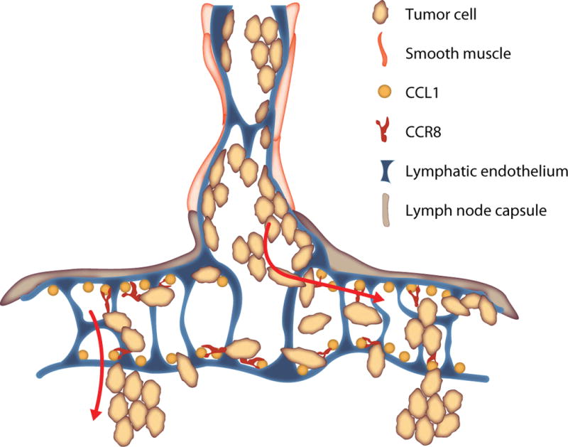
In cancer, lymphatic vasculature has been traditionally viewed only as a transportation system for metastatic cells. It has now become clear that lymphatics perform many additional functions which could influence cancer progression. Lymphangiogenesis, induced at the primary tumor site and at distant sites, potently augments metastasis. Lymphatic endothelial cells (LECs) control tumor cell entry and exit from the lymphatic vessels. LECs also control immune cell traffic and directly modulate adaptive immune responses. This review highlights advances in our understanding of the mechanisms by which lymphatic vessels, and in particular lymphatic endothelium, impact metastasis.
Keywords: CCL1; CCR8; Chemokine; Immunoregulation; Lymph node; Lymphangiogenesis; Lymphatic endothelium; Metastasis; VEGF-C; VEGFR-3.
Copyright © 2014 Elsevier Inc. All rights reserved.
Figures
References
-
- Acikgoz G, Kim SM, Houseni M, Cermik TF, Intenzo CM, Alavi A. Pulmonary lymphangitic carcinomatosis (PLC): spectrum of FDG-PET findings. Clin Nucl Med. 2006;31:673–678. - PubMed
-
- Alitalo K, Carmeliet P. Molecular mechanisms of lymphangiogenesis in health and disease. Cancer Cell. 2002;1:219–227. - PubMed
-
- Amundson DE, Weiss PJ. Hypoxemia in malignant carcinoid syndrome: a case attributed to occult lymphangitic metastatic involvement. Mayo Clin Proc. 1991;66:1178–1180. - PubMed
-
- Angeli V, Ginhoux F, Llodra J, Quemeneur L, Frenette PS, Skobe M, Jessberger R, Merad M, Randolph GJ. B cell-driven lymphangiogenesis in inflamed lymph nodes enhances dendritic cell mobilization. Immunity. 2006;24:203–215. - PubMed
-
- Ben-Baruch A. Organ selectivity in metastasis: regulation by chemokines and their receptors. Clin Exp Metastasis. 2008;25:345–356. - PubMed
Publication types
MeSH terms
Grants and funding
LinkOut - more resources
Full Text Sources
Other Literature Sources
