. 2014 Dec 15;23(24):6644-58.
doi: 10.1093/hmg/ddu372. Epub 2014 Jul 15.

SUCLG2 identified as both a determinator of CSF Aβ1-42 levels and an attenuator of cognitive decline in Alzheimer's disease

Affiliations

SUCLG2 identified as both a determinator of CSF Aβ1-42 levels and an attenuator of cognitive decline in Alzheimer's disease

Alfredo Ramirez et al. Hum Mol Genet. .

Abstract

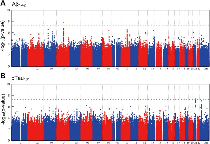
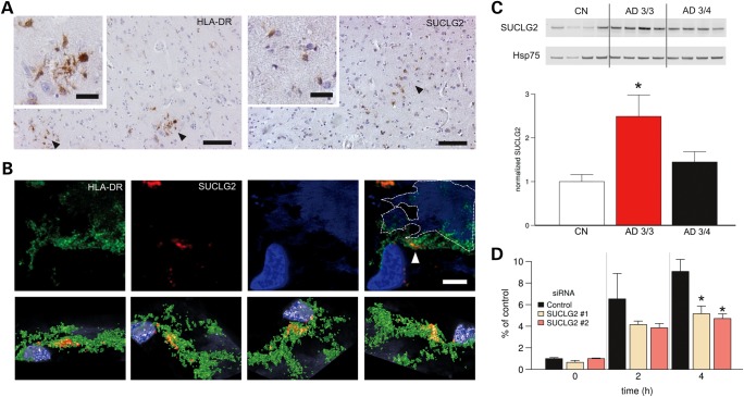
Cerebrospinal fluid amyloid-beta 1-42 (Aβ1-42) and phosphorylated Tau at position 181 (pTau181) are biomarkers of Alzheimer's disease (AD). We performed an analysis and meta-analysis of genome-wide association study data on Aβ1-42 and pTau181 in AD dementia patients followed by independent replication. An association was found between Aβ1-42 level and a single-nucleotide polymorphism in SUCLG2 (rs62256378) (P = 2.5×10(-12)). An interaction between APOE genotype and rs62256378 was detected (P = 9.5 × 10(-5)), with the strongest effect being observed in APOE-ε4 noncarriers. Clinically, rs62256378 was associated with rate of cognitive decline in AD dementia patients (P = 3.1 × 10(-3)). Functional microglia experiments showed that SUCLG2 was involved in clearance of Aβ1-42.


Figures

Figure 1.
Figure 1.
Genome-wide signal intensity (Manhattan) plots. (A) Results for the association with CSF Aβ1–42. (B) Results for the association with CSF pTau181. Both plots show the individual P-values against genomic position. Chromosome number is shown on the x-axis. The results within each chromosome are plotted left to right, with left being the P-terminal end of the corresponding chromosome. Horizontal dashed lines indicate a P-value threshold of 5 × 10⁻⁸ (i.e. genome-wide significance). Green dots depict single-nucleotide polymorphisms with genome-wide significance in the genome-wide association study step.
Figure 2.
Figure 2.
Functional studies of SUCLG2. (A and B) SUCLG2 localized to microglia in AD brains. (A) Representative DAB immunostaining with SUCLG2 and HLA-DR (as a human microglial marker) in brain cortex of AD subjects. DAB-positive cells with characteristic romboidal nuclei (typically microglial cells) are indicated by arrows. Scale bars are 100 and 20 µm on the main and small images, respectively. (B) High-resolution confocal image of an area of frontal cortex from an AD dementia patient immune labelled with HLA-DR (green) and SUCLG2 (red). Plaque autofluorescence is visible in the blue channel and it is highlighted with a discontinuous line. Most of the SUCLG2 is enclosed by the HLA-DR (arrow). The top panel shows several viewpoints of a 3D reconstruction of the same area, which is shown in the lower panel. (Scale is 10 μm.) (C) Increased levels of SUCLG2 in AD ApoE 3/3 brains. Soluble extracts from postmortem frontal cortex of AD dementia patients with ApoE 3/4 and ApoE 3/3 status plus age-matched healthy controls (CN) were probed with an antibody to SUCLG2. Expression levels were normalized to Hsp75 (CN, n = 6; AD, 3/4, n = 8; AD, 3/3, n = 4). Data are presented as mean ± SEM. Asterisk denotes P < 0.05. (D) SUCLG2 silencing is detrimental to extracellular Aβ1–42. FAM-Aβ1–42 phagocytosis was impaired in BV2 cells in which SUCLG2 was silenced by two distinct siRNA compared with negative control siRNA (n = 6). FAM-Aβ1–42 was added at a 500 nm final concentration and the amount of internalized Aβ1–42 was assessed at the indicated time points, showing that cells contain less Aβ1–42 in the absence of SUCLG2. Data are presented as mean ± SEM. Asterisk denotes P < 0.05.

References

    1. Kim S., Swaminathan S., Shen L., Risacher S.L., Nho K., Foroud T., Shaw L.M., Trojanowski J.Q., Potkin S.G., Huentelman M.J., et al. Genome-wide association study of CSF biomarkers Abeta1–42, t-tau, and p-tau181p in the ADNI cohort. Neurology. 2011;76:69–79. - PMC - PubMed
    1. Schjeide B.M., Schnack C., Lambert J.C., Lill C.M., Kirchheiner J., Tumani H., Otto M., Tanzi R.E., Lehrach H., Amouyel P., et al. The role of clusterin, complement receptor 1, and phosphatidylinositol binding clathrin assembly protein in Alzheimer disease risk and cerebrospinal fluid biomarker levels. Arch. Gen. Psychiatry. 2011;68:207–213. - PubMed
    1. Elias-Sonnenschein L.S., Helisalmi S., Natunen T., Hall A., Paajanen T., Herukka S.K., Laitinen M., Remes A.M., Koivisto A.M., Mattila K.M., et al. Genetic loci associated with Alzheimer's disease and cerebrospinal fluid biomarkers in a Finnish case-control cohort. PLoS ONE. 2013;8:e59676. - PMC - PubMed
    1. Han M.R., Schellenberg G.D., Wang L.S. Genome-wide association reveals genetic effects on human Abeta42 and tau protein levels in cerebrospinal fluids: a case control study. BMC Neurol. 2010;10:90. - PMC - PubMed
    1. Cruchaga C., Kauwe J.S., Harari O., Jin S.C., Cai Y., Karch C.M., Benitez B.A., Jeng A.T., Skorupa T., Carrell D., et al. GWAS of cerebrospinal fluid tau levels identifies risk variants for Alzheimer's disease. Neuron. 2013;78:256–268. - PMC - PubMed

Publication types

MeSH terms