. 2014 Dec;22(12):2118-2129.
doi: 10.1038/mt.2014.133. Epub 2014 Jul 16.

A cationic nanoemulsion for the delivery of next-generation RNA vaccines

Affiliations

A cationic nanoemulsion for the delivery of next-generation RNA vaccines

Luis A Brito et al. Mol Ther. 2014 Dec.

Abstract

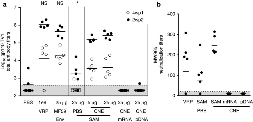
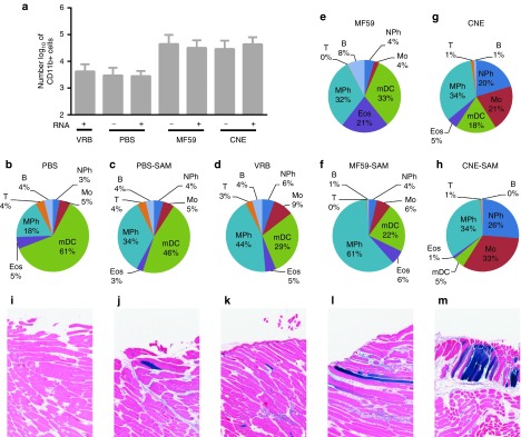
Nucleic acid-based vaccines such as viral vectors, plasmid DNA, and mRNA are being developed as a means to address a number of unmet medical needs that current vaccine technologies have been unable to address. Here, we describe a cationic nanoemulsion (CNE) delivery system developed to deliver a self-amplifying mRNA vaccine. This nonviral delivery system is based on Novartis's proprietary adjuvant MF59, which has an established clinical safety profile and is well tolerated in children, adults, and the elderly. We show that nonviral delivery of a 9 kb self-amplifying mRNA elicits potent immune responses in mice, rats, rabbits, and nonhuman primates comparable to a viral delivery technology, and demonstrate that, relatively low doses (75 µg) induce antibody and T-cell responses in primates. We also show the CNE-delivered self-amplifying mRNA enhances the local immune environment through recruitment of immune cells similar to an MF59 adjuvanted subunit vaccine. Lastly, we show that the site of protein expression within the muscle and magnitude of protein expression is similar to a viral vector. Given the demonstration that self-amplifying mRNA delivered using a CNE is well tolerated and immunogenic in a variety of animal models, we are optimistic about the prospects for this technology.


Figures

Figure 1
Figure 1
Physical characterization of cationic nanoemulsion (CNE)-formulated SAM. (a) Schematic illustration and role of components of CNE in complex with SAM RNA. (b) Cryo-EM of CNE prior to RNA addition. (c) Particle size measured by dynamic light scattering of CNE before (red line) and after (green line) SAM RNA addition. Data are reported as the Z-average (Z-ave) with the poly-dispersity index (pdi). (d) Denaturing RNA agarose gel electrophoresis showing protection of SAM RNA from RNAse: molecular weight ladder (lane 1), SAM RNA (lane 2), SAM RNA after incubation with RNase (lane 3), SAM RNA after extraction from CNE (lane 4), CNE with SAM RNA after RNase exposure, inactivation of RNase, and RNA extraction from CNE (lane 5).
Figure 2
Figure 2
Immunogenicity of RSV SAM vaccine and comparators in mice. Groups of eight BALB/c mice were immunized i.m. on days 0 and 21 with 3 µg RSV-F subunit antigen with MF59, 1 × 106 IU RSV F-expressing VRP, 15 µg SAM RNA expressing RSV F in phosphate-buffered saline (PBS), 15 µg mRNA expressing RSV F in PBS, 15 µg pDNA expressing RSV F in PBS, 0.015 µg SAM RNA expressing RSV F formulated with cationic nanoemulsion (CNE), 0.15 µg SAM RNA expressing RSV F formulated with CNE, 1.5 µg SAM RNA expressing RSV F formulated with CNE, 15 µg SAM RNA expressing RSV F formulated with CNE, 1.5 µg mRNA expressing RSV F formulated with CNE, 15 µg mRNA expressing RSV F formulated with CNE, 1.5 µg pDNA expressing RSV F formulated with CNE, and 15 µg pDNA expressing RSV F formulated with CNE. Control mice were not injected with any formulation. Sera were collected 2 weeks after the second vaccination. (a) F-specific IgG titers measured by enzyme-linked immunosorbent assay (ELISA). Data are from individual mice (depicted as dots), and the geometric mean titers (GMTs) are solid lines. (b) RSV 60% neutralization titers 2 weeks after the second vaccination. Data are from pooled sera (two pools per group, depicted as dots), and the GMTs as solid lines. The dotted lines indicate assay limits of detection (titers of 25 for ELISA, 20 for neutralizing antibodies). For calculation of GMTs, titers below the limits of detection were assigned a value of 5 for ELISA and 10 for neutralization. NS, not statistically different from 15 µg SAM RNA expressing RSV F formulated with CNE (analysis of variance (ANOVA), Tukey's post-test), * statistically different from 15 µg SAM RNA expressing RSV F formulated with CNE (P = 0.001 ANOVA, Tukey's post-test).
Figure 3
Figure 3
Immunogenicity of HIV SAM vaccine and comparators in rabbits. Groups of five New Zealand white rabbits were immunized i.m. on days 0 and 21 with PBS, 1 × 108 IU HIV gp140 expressing VRP, 25 µg HIV gp140 env with MF59, 25 µg SAM RNA expressing HIV gp140 in PBS, 5 µg SAM RNA expressing HIV gp140 formulated with cationic nanoemulsion (CNE), 25 µg SAM RNA expressing HIV gp140 formulated with CNE, 25 µg mRNA expressing HIV gp140 formulated with CNE, or 25 µg pDNA expressing HIV gp140 formulated with CNE. Sera were collected 4 weeks post first (4wp1) immunization (open circles) and 2 weeks post second (2wp2) immunization (closed circles). (a) gp140-specific IgG titers measured by enzyme-linked immunosorbent assay (ELISA). Data are from individual rabbits (depicted as dots), and the geometric mean titers (GMTs) are solid lines. (b) HIV MW965.26 serum neutralizing antibody titers elicited by 1 × 108 IU HIV gp140 expressing VRP, 25 µg SAM RNA expressing HIV gp140 in PBS, 25 µg SAM RNA expressing HIV gp140 formulated with CNE, 25 µg mRNA expressing HIV gp140 formulated with CNE, and 25 µg pDNA expressing HIV gp140 formulated with CNE. Data are from the individual sera (depicted as dots) tested 2 weeks after the second vaccination, and the GMT is a solid line. The dotted lines indicate assay limits of detection (400 for ELISA, 20 for neutralization). For calculation of GMTs, titers below the limit of detections were assigned a value of 200 for ELISA and 10 for neutralization. NS, not statistically different from 25 µg SAM RNA expressing HIV gp140 formulated with CNE (analysis of variance (ANOVA), Tukey's post-test), * statistically different from 15 µg SAM RNA expressing RSV F formulated with CNE (P = 0.001 ANOVA, Tukey's post-test).
Figure 4
Figure 4
Immunogenicity in rhesus macaques. A group of six rhesus macaques was immunized intramuscularly on days 0 and 28 with 75 µg SAM RNA expressing hCMV gB formulated with cationic nanoemulsion (CNE) and 75 µg of SAM RNA expressing hCMV pp65-IE1 formulated with CNE. (a) gB-specific IgG titers measured by enzyme-linked immunosorbent assay (ELISA). Data are presented as geometric mean titers (GMTs) ± standard error of the mean (SEM) 2 weeks post first immunization (2wp1), 2 weeks post second immunization (2wp2), and 8 weeks post second immunization (8wp2). (b) CMV clinical isolate 8819 neutralization titers on ARPE-19 cells; data are presented as GMTs ± SEM, 2wp1 immunization, 2wp2 immunization, and 8wp2 immunization. The dotted lines indicate preimmunization titers (2,381 for gB ELISA, 422 for CMV neutralization).
Figure 5
Figure 5
Circulating antigen-specific T cells in immunized rhesus macaques. Blood was drawn and peripheral blood monocytes (PBMCs) were cryopreserved from six rhesus macaques that were immunized i.m. on days 0 and 28 with 75 µg SAM RNA expressing hCMV gB formulated with cationic nanoemulsion (CNE) and 75 µg of SAM RNA expressing hCMV pp65-IE1 formulated with CNE (same animals as Figure 4). PBMCs were thawed, stimulated, and analyzed as described in Materials and Methods. PBMCs were available for five of the six rhesus. Percentages of cytokine + antigen-specific CD4+ and CD8− T cells in individual rhesus after stimulation in vitro with a hCMV gB peptide mix (black checkered), hCMV pp65 peptide mix (black), or hCMV IE-1 peptide mix (gray). (a) 21 days prior to the first immunization and (b) 14 days after the second immunization. Percentages of antigen-specific CD8+ CD4− T cells in individual rhesus after stimulation in vitro with a CMV gB peptide mix (black checkered), CMV pp65 peptide mix (black), or CMV IE-1 peptide mix (gray) (c) 21 days prior to the first immunization and (d) 14 days after the second immunization.
Figure 6
Figure 6
Luciferase activity in mice. Groups of five BALB/c mice were inoculated i.m. on day 0 with either phosphate-buffered saline (PBS) (closed grey diamond), 5 × 106 IU luc expressing VRP (open black diamond), 1.5 µg SAM RNA expressing luciferase formulated with CNE at a 1.5 dose (open dark gray circle) or 15 µg dose (closed dark gray circle), mRNA expressing luciferase formulated with CNE at a 1.5 µg dose (open gray square) or 15 µg dose (closed gray square), or pDNA expressing luciferase formulated with CNE at a 1.5 µg dose (open light gray triangle) or 15 µg dose (closed light gray triangle). Average total flux (photons/second) of bioluminescence emanating from the injection site was measured repeatedly over the course of 10 weeks. Total flux throughout the whole 10-week experiment (a), total flux throughout the first 5 days of the experiment (b), peak expression as measured by total flux, time of peak expression is noted in parenthesis (c).
Figure 7
Figure 7
Antigen expression, localization, and cell infiltration. BALB/c mice were inoculated i.m. with phosphate-buffered saline (PBS) (n = 12), 15 µg SAM RNA expressing green fluorescent protein (GFP) in PBS (n = 4), 5 × 106 IU VRP expressing GFP (n = 4), MF59 (n = 8), 15 µg SAM RNA expressing GFP formulated with MF59 (n = 8), cationic nanoemulsion (CNE) (n = 8), or 15 µg SAM RNA expressing GFP formulated with CNE (n = 8). (a) CD11b+ cells found in collagenase dissociated muscle cell suspensions 3 days after immunization. (b–h) Leukocyte infiltrate composition 3 days after administration of (b,c) PBS (−/+ 15 µg SAM RNA expressing GFP), (d) 5 × 106 IU VRP expressing GFP, (e,f) MF59 (−/+ 15 µg SAM RNA expressing GFP), (g,h) CNE (−/+ 15 µg SAM RNA expressing GFP); B, B-cell; Eos, eosinophil; Mo, monocyte; mDC, dendritic cell; MPh, macrophage; NPh, neutrophil; T, T-cell. (i–m) Representative histological tissue sections stained for β-gal 3 days postinnoculation of BALB/c mice after administration with (i) PBS, (j) 15 µg SAM RNA expressing β-gal formulated with PBS, (k) 1 × 106 IU VRP expressing β-gal, (l) 15 µg SAM RNA expressing β-gal formulated with MF59, (m) 15 µg SAM RNA expressing β-gal formulated with CNE.
Figure 8
Figure 8
Comparison of immunogenicity of SAM RNA delivered with MF59 or cationic nanoemulsion (CNE). Groups of 8 BALB/c mice were immunized i.m. on days 0 and 21 with 1 µg SAM RNA expressing RSV F in phosphate-buffered saline (PBS), 1 µg SAM RNA expressing RSV F formulated with MF59, or 1 µg SAM RNA expressing RSV F formulated with CNE. Sera were collected two weeks after the second vaccination. F-specific serum IgG titers measured by enzyme-linked immunosorbent assay (ELISA). Data are from individual mice (depicted as dots), and the geometric mean titers (GMTs) are solid lines. Error bars depict the standard error of the mean. * statistically different from RSV F SAM RNA in PBS and SAM RNA expressing RSV F formulated with MF59 (P = 0.001 analysis of variance, Tukey's post-test).

References

    1. Rappuoli R, Mandl CW, Black S, De Gregorio E. Vaccines for the twenty-first century society. Nat Rev Immunol. 2011;11:865–872. - PMC - PubMed
    1. Kutzler MA, Weiner DB. DNA vaccines: ready for prime time. Nat Rev Genet. 2008;9:776–788. - PMC - PubMed
    1. Sardesai NY, Weiner DB. Electroporation delivery of DNA vaccines: prospects for success. Curr Opin Immunol. 2011;23:421–429. - PMC - PubMed
    1. Geall AJ, Mandl CW, Ulmer JB. RNA: The new revolution in nucleic acid vaccines. Seminars in Immunol. 2013;25:152–159. - PubMed
    1. Geall AJ, Verma A, Otten GR, Shaw CA, Hekele A, Banerjee K, et al. Nonviral delivery of self-amplifying RNA vaccines. Proc Natl Acad Sci USA. 2012;109:14604–14609. - PMC - PubMed

Publication types