Review
. 2014:385:327-56.
doi: 10.1007/82_2014_394.

Secondary bacterial infections in influenza virus infection pathogenesis

Affiliations
Review

Secondary bacterial infections in influenza virus infection pathogenesis

Amber M Smith et al. Curr Top Microbiol Immunol. 2014.

Abstract

Influenza is often complicated by bacterial pathogens that colonize the nasopharynx and invade the middle ear and/or lung epithelium. Incidence and pathogenicity of influenza-bacterial coinfections are multifactorial processes that involve various pathogenic virulence factors and host responses with distinct site- and strain-specific differences. Animal models and kinetic models have improved our understanding of how influenza viruses interact with their bacterial co-pathogens and the accompanying immune responses. Data from these models indicate that considerable alterations in epithelial surfaces and aberrant immune responses lead to severe inflammation, a key driver of bacterial acquisition and infection severity following influenza. However, further experimental and analytical studies are essential to determining the full mechanistic spectrum of different viral and bacterial strains and species and to finding new ways to prevent and treat influenza-associated bacterial coinfections. Here, we review recent advances regarding transmission and disease potential of influenza-associated bacterial infections and discuss the current gaps in knowledge.


Figures

Fig. 1
Fig. 1
Animal models of influenza-associated bacterial coinfections. Several different animal models have been used to study the effect that influenza viruses have on bacterial transmission and colonization and on invasive diseases, such as acute otitis media and pneumonia (Wherry and Butterfield ; Shope ; Francis and de Torregrosa ; Berendt et al. ; Rarey et al. ; Hajek et al. ; Hirano et al. ; McCullers and Rehg ; Okamoto et al. ; Seki et al. ; Peltola et al. ; Montgomery et al. ; Small et al. ; Diavatopoulos et al. ; Lee et al. ; Jamieson et al. ; McCullers et al. ; Loving et al. ; Iverson et al. ; Kudva et al. ; Ayala et al. ; Chaussee et al. ; Short et al. ; Mina et al. ; McHugh et al. ; Redford et al. 2014)
Fig. 2
Fig. 2
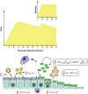
Influenza-bacterial interaction during coinfections. Numerous alterations of the respiratory epithelium and host immune responses occur during influenza virus infection that predisposes a host to coinfection with bacterial pathogens. As influenza virus infects and kills host cells, epithelial surfaces become exposed and permissive to bacterial attachment. Physical barriers (e.g., mucociliary transport) are damaged, pathogen detection is decreased, anti-microbial peptides (AMPs) are downregulated, receptors are upregulated, virus production is enhanced, bacterial transepithelial migration is permitted, and repair mechanisms are lost. Several host responses are also dampened, altered, or removed. Alveolar macrophages, neutrophils, dendritic cells, and NK cells have altered cytokine profiles and become impaired and/or depleted. These changes result in a heightened inflammatory environment with decreased bacterial surveillance and eradication
Fig. 3
Fig. 3
Kinetics of influenza-pneumococcal coinfection. Model schematic and equations that result in the observed kinetics of influenza virus infection followed by pneumococcus given 7 days postinfluenza infection (Smith et al. 2013). During primary influenza, susceptible epithelial (target) cells (T) become infected at a rate βV per cell. Infected cells (I 1) first undergo an eclipse phase at rate k per cell prior to entering a state (I 2) in which virus is produced. Productively infected cells are lost, through apoptosis, viral cytopathic effects, or removal by immune cells, at a rate δ per cell. Virus (V) is produced at rate p per cell, which is significantly increased by bacterial presence (aP z) (boxed), and cleared at rate c. Invading pneumococci (P) proliferate at maximum rate r with a tissue capacity K p CFU/ml. Bacteria are cleared via phagocytosis by alveolar macrophages (MA) at rate γf per cell, which is significantly reduced by virus presence formula image (boxed). With this kinetic description, viral titers increase exponentially, peak and begin to decline prior to bacterial invasion. Once bacteria are present, a viral rebound occurs and bacteria grow exponentially before reaching a maximum capacity. The potential increase in bacterial adherence to virus-infected cells and any accompanying cell death has little effect are excluded here

References

    1. Abramson JS, Giebink GS, Mills EL, Quie PG. Polymorphonuclear leukocyte dysfunction during influenza virus infection in chinchillas. J Infect Dis. 1981;143:836–845. - PubMed
    1. Abramson JS, Giebink GS, Quie PG. Influenza A virus-induced polymorphonuclear leukocyte dysfunction in the pathogenesis of experimental pneumococcal otitis media. Infect Immun. 1982;36:289–296. - PMC - PubMed
    1. Abt MC, Osborne LC, Monticelli LA, et al. Commensal bacteria calibrate the activation threshold of innate antiviral immunity. Immunity. 2012;37:158–170. - PMC - PubMed
    1. Alymova IV, Green AM, van de Velde N, et al. Immunopathogenic and antibacterial effects of H3N2 influenza A virus PB1-F2 map to amino acid residues 62, 75, 79, and 82. J Virol. 2011;85:12324–12333. - PMC - PubMed
    1. Ansaldi F, de Florentiis D, Parodi V, et al. Bacterial carriage and respiratory tract infections in subjects > or = 60 years during an influenza season: implications for the epidemiology of community acquired pneumonia and influenza vaccine effectiveness. J Prev Med Hyg. 2012;53:94–97. - PubMed

Publication types

MeSH terms