Review
. 2014 Aug;15(8):839-52.
doi: 10.15252/embr.201439076. Epub 2014 Jul 15.

Getting ready for building: signaling and autophagosome biogenesis

Affiliations
Review

Getting ready for building: signaling and autophagosome biogenesis

Adi Abada et al. EMBO Rep. 2014 Aug.

Abstract

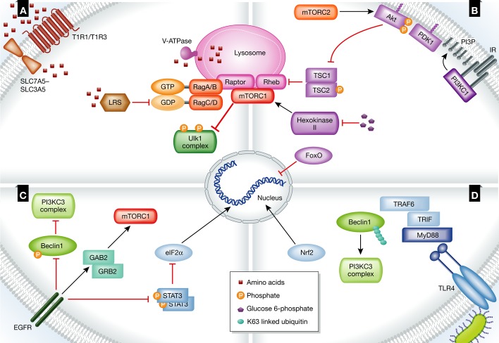
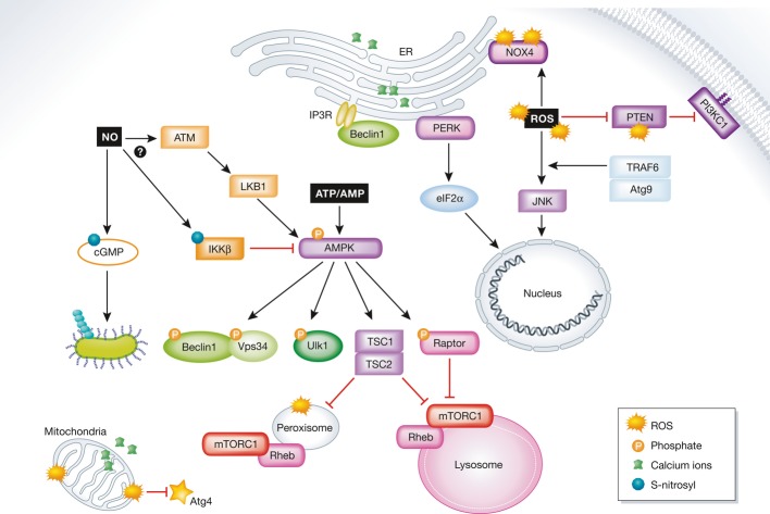
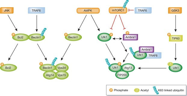
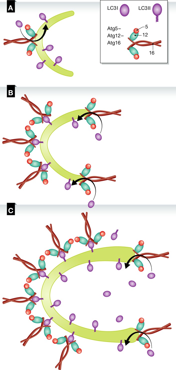
Autophagy is the main cellular catabolic process responsible for degrading organelles and large protein aggregates. It is initiated by the formation of a unique membrane structure, the phagophore, which engulfs part of the cytoplasm and forms a double-membrane vesicle termed the autophagosome. Fusion of the outer autophagosomal membrane with the lysosome and degradation of the inner membrane contents complete the process. The extent of autophagy must be tightly regulated to avoid destruction of proteins and organelles essential for cell survival. Autophagic activity is thus regulated by external and internal cues, which initiate the formation of well-defined autophagy-related protein complexes that mediate autophagosome formation and selective cargo recruitment into these organelles. Autophagosome formation and the signaling pathways that regulate it have recently attracted substantial attention. In this review, we analyze the different signaling pathways that regulate autophagy and discuss recent progress in our understanding of autophagosome biogenesis.

Keywords: Atgs; autophagosome biogenesis; autophagy; mTOR; signaling.


Figures

Figure 1
Figure 1. Regulation of autophagy by extracellular cues
(A) Amino acids are key regulators of autophagy. When they are in excess, mTORC1 is targeted to the lysosomal membrane, where it is activated by Rheb and inhibits autophagy through phosphorylation of Ulk1 complex subunits. (B) Binding of insulin to its receptor (IR) activates mTOR via the PI3KC1/Akt/TSC pathway, inhibiting autophagy. The expression of autophagy-related proteins is inhibited after the inhibition of FoxO transcription factors by Akt. Glucose 6-phosphate inhibits the activity of hexokinase-II, an mTOR activator, inhibiting autophagy. (C) Activation of EGFR by its ligand inhibits autophagy directly by the phosphorylation of Beclin1 or indirectly via GRB2 and GAB2, as well as via the phosphorylation of STAT3, which releases eIF2α to induce the expression of autophagy-related proteins. (D) TLR4 is activated upon binding of LPS, leading to the recruitment of adaptor proteins to the plasma membrane. As a consequence, TRAF6 is recruited, resulting in the Lys63-linked ubiquitination of Beclin1, allowing it to bind PI3KC3 and induce autophagy. Nrf2 is activated, up-regulating the expression of p62. See Glossary for definitions and the text for details.
Figure 2
Figure 2. Regulation of autophagy by intracellular cues
Internal cues regulate autophagy on different levels from many intracellular locations. The activity of mTORC1 is regulated at the lysosome and the peroxisome through AMPK. Active AMPK indirectly inhibits autophagy by activating the TSC1/2 complex and via inhibition of raptor by phosphorylation, both of which lead to the inhibition of mTORC1. Autophagy is inhibited directly by Ulk1, Vps34, and Beclin1 phosphorylation. ROS molecules activate autophagy at the plasma membrane, the ER, and mitochondria, as well as by up-regulating the expression of autophagy-related proteins. Ca2+ signaling is mediated from its intracellular storages in the mitochondria and the ER. The regulation of autophagy through AMPK induced by NO remains poorly understood. NO regulates mitophagy through cGMP. See Glossary for definitions and the text for details.
Figure 3
Figure 3. Post-translational modifications regulate PI3KC3 and the Ulk1 complex
Ubiquitination, phosphorylation, and acetylation of Ulk1 and Beclin1 regulate autophagy by promoting or preventing the formation of the Ulk1 complex or PI3KC3. See Glossary for definitions and the text for details.
Figure 4
Figure 4. Model of the Atg12–Atg5–Atg16 complex function in autophagy
(A) The Atg12–Atg5–Atg16 complex is recruited to the phagophore after its initial nucleation. At this stage, the membrane is curved and the complex promotes the lipidation of LC3 with PE. (B) Once the membrane elongates, the complex remains associated with the membrane through LC3 on membranes with low curvature for their stabilization and continues to promote LC3 lipidation at the highly curved edges of the phagophore. (C) As the elongation continues, the Atg12–Atg5–Atg16 complex, together with the lipid-conjugated LC3, forms a coat-like structure that stabilizes the structure of the phagophore.
Figure 5
Figure 5. Models of autophagosome biogenesis
(A) The current view of autophagosome biogenesis is a continuous process, where an initial membrane for autophagosome formation buds out from an existing organelle and is further elongated by the fusion of vesicles, some containing Atg9. The Atg12–Atg5–Atg16 complex promotes LC3 lipidation on the highly curved membranes while supporting the membrane’s structure as it elongates. Once the autophagosome is sealed, it encapsulates cargo for degradation and the external biogenesis machinery is removed. (B) A new model for autophagosome biogenesis. Multiple nucleation membranes bud from several organelles to contribute to the formation of the initial membrane of the autophagosome. Each membrane elongates individually until all are fused to create an autophagosome that encapsulates cargo for lysosomal degradation.

References

    1. Boya P, Reggiori F, Codogno P. Emerging regulation and functions of autophagy. Nat Cell Biol. 2013;15:713–720. - PMC - PubMed
    1. Choi AM, Ryter SW, Levine B. Autophagy in human health and disease. N Engl J Med. 2013;368:651–662. - PubMed
    1. Sato M, Sato K. Dynamic regulation of autophagy and endocytosis for cell remodeling during early development. Traffic. 2013;14:479–486. - PubMed
    1. Guan JL, Simon AK, Prescott M, Menendez JA, Liu F, Wang F, Wang C, Wolvetang E, Vazquez-Martin A, Zhang J. Autophagy in stem cells. Autophagy. 2013;9:830–849. - PMC - PubMed
    1. Weidberg H, Shvets E, Elazar Z. Biogenesis and cargo selectivity of autophagosomes. Annu Rev Biochem. 2011;80:125–156. - PubMed

Publication types

Substances

LinkOut - more resources