. 2014 Jul 15;5(4):e01188-14.
doi: 10.1128/mBio.01188-14.

Toxoplasma gondii ingests and digests host cytosolic proteins

Affiliations

Toxoplasma gondii ingests and digests host cytosolic proteins

Zhicheng Dou et al. mBio. .

Abstract

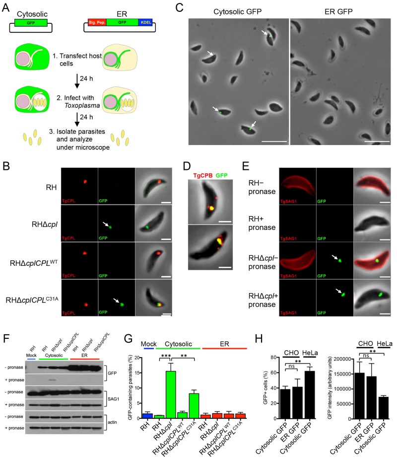
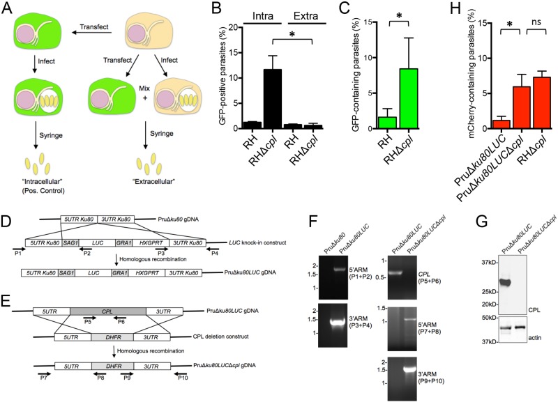
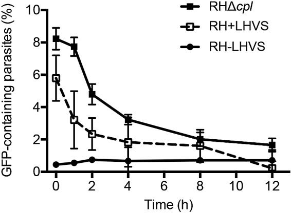
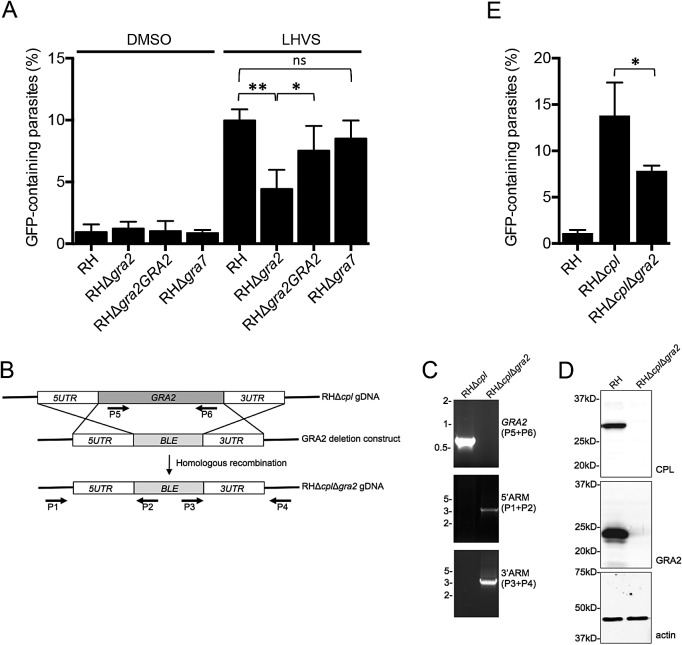
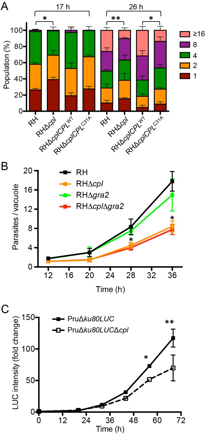
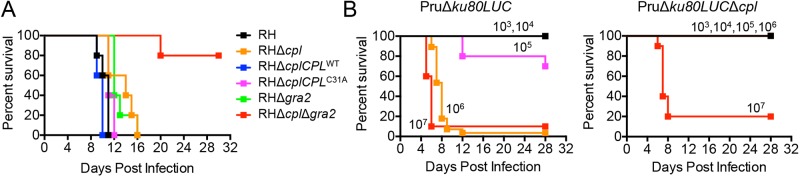
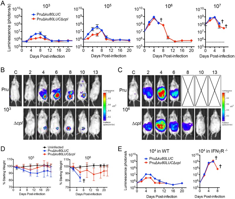
The protozoan parasite Toxoplasma gondii resides within a nonfusogenic vacuole during intracellular replication. Although the limiting membrane of this vacuole provides a protective barrier to acidification and degradation by lysosomal hydrolases, it also physically segregates the parasite from the host cytosol. Accordingly, it has been suggested that T. gondii acquires material from the host via membrane channels or transporters. The ability of the parasite to internalize macromolecules via endocytosis during intracellular replication has not been tested. Here, we show that Toxoplasma ingests host cytosolic proteins and digests them using cathepsin L and other proteases within its endolysosomal system. Ingestion was reduced in mutant parasites lacking an intravacuolar network of tubular membranes, implicating this apparatus as a possible conduit for trafficking to the parasite. Genetic ablation of proteins involved in the pathway is associated with diminished parasite replication and virulence attenuation. We show that both virulent type I and avirulent type II strain parasites ingest and digest host-derived protein, indicating that the pathway is not restricted to highly virulent strains. The findings provide the first definitive evidence that T. gondii internalizes proteins from the host during intracellular residence and suggest that protein digestion within the endolysosomal system of the parasite contributes to toxoplasmosis. Importance: Toxoplasma gondii causes significant disease in individuals with weak immune systems. Treatment options for this infection have drawbacks, creating a need to understand how this parasite survives within the cells it infects as a prelude to interrupting its survival strategies. This study reveals that T. gondii internalizes proteins from the cytoplasm of the cells it infects and degrades such proteins within a digestive compartment within the parasite. Disruption of proteins involved in the pathway reduced parasite replication and lessened disease severity. The identification of a novel parasite ingestion pathway opens opportunities to interfere with this process and improve the outcome of infection.


Figures

FIG 1
FIG 1
Experimental design and discovery of the ingestion pathway. (A) Experimental design with GFP constructs expressing cytosolic or ER GFP. Constructs were transiently transfected into CHO cells, and expression was allowed to develop for 24 h before infection with the indicated strains. Parasites were mechanically liberated from host cells after 24 h of replication and examined by fluorescence microscopy. (B) Host cytosolic GFP is associated with RHΔcpl and RHΔcplCPLC31A strains lacking cathepsin L activity but not WT or genetically wild-type CPL-complemented parasites. Mechanically liberated parasites were fixed and stained with antibodies to CPL. Bar, 2 µm. (C) Fluorescent overlay images of RHΔcpl parasites harvested from host cells expressing the indicated GFP. Bars, 10 µm. (D) GFP colocalizes with CBP in the VAC of a subset of parasites. Shown are two examples of GFP-positive parasites stained with anti-CPB. Bars, 10 µm. (E) GFP is shielded from pronase treatment in RHΔcpl parasites. Parasites harvested from GFP-expressing host cells were treated with pronase to test for protease protection as an indicator of internalization. Parasites were stained with antibodies to the surface antigen SAG1, which was proteolysed on treated parasites. Bars, 2 µm. (F) Quantification of GFP in the indicated strains liberated from host cells transfected with constructs for expression of cytosolic or ER GFP. Statistical significance by unpaired Student’s t test: **, P < 0.01; ***, P < 0.001. (G) Immunoblot detection of ingested GFP after pronase treatment. RH, RHΔcpl, and RHΔcplCPLWT parasites were used to infect CHO cells transiently expressing cytosolic and ER GFP proteins, respectively. Twenty-four hours postinfection, parasites were harvested and treated with pronase and saponin. The lysates of treated parasites were probed with anti-GFP antibody. Lysates were also probed with polyclonal antibodies against actin and SAG1 as a loading control and by measurement of pronase digestion, respectively. (H) Transfection efficiency of cytosolic and ER-retained GFP in CHO and HeLa cells. DNA constructs were transfected into CHO and HeLa cells. Twenty-four hours posttransfection, cells were trypsinized and analyzed by flow cytometry to determine the percentage of GFP-positive cells (left) and GFP intensity (right). Values shown in panels A and B are means ± standard deviations (SD) from n = 3 experiments. Significance by unpaired Student’s t test: *, P < 0.05; **, P < 0.01. ns, not significant (i.e., P > 0.05).
FIG 2
FIG 2
GFP is ingested during intracellular replication. (A) Experimental scheme of testing for the absence of GFP ingestion during parasite harvest. As a positive (Pos.) control (left side of the scheme), CHO cells were transfected with the cytosolic GFP expression construct, infected 24 h posttransfection with RH or RHΔcpl parasites, and mechanically liberated by syringing 24 h postinfection. These were termed “intracellular” parasites. To test for uptake of host cytosolic GFP during mechanical rupture and harvest (right side of the scheme), CHO cells were transiently transfected and mixed with untransfected and infected CHO cells, and parasites were mechanically liberated by syringing. These were termed “extracellular” parasites because internalization could only occur after the parasites were liberated. (B) GFP is not ingested during harvest, indicating ingestion during intracellular replication. Shown is the percentage of GFP-positive parasites harvested by the “intracellular” (intra) or “extracellular” (extra) scheme. Statistical significance by unpaired Student’s t test: *, P < 0.05. (C) GFP transiently expressed in HeLa cells is associated with CPL-deficient parasites, indicating that the parasites also ingest host cytosolic proteins from human cells. Statistical significance by unpaired Student’s t test: *, P < 0.05. (D) Schematic illustration of the strategy for creating PruΔku80LUC parasites. A luciferase expression cassette (LUC) was transfected into PruΔku80 parasites and selected with mycophenolic acid and xanthine for double crossover insertion via homologous recombination at the “empty” ku80 locus. (E) A CPL deletion construct was transfected into PruΔku80LUC parasites and selected with pyrimethamine for double crossover replacement of the CPL by homologous recombination. (F) Primers indicated in panels D and E were used to verify the insertion of LUC at the KU80 locus and CPL deletion by PCR and agarose gel electrophoresis. ARM, the end of the target gene. (G) Cell lysates were immunoblotted with antibodies against CPL to confirm the knockout and with antibodies for actin as a loading control. (H) PruΔku80LUCΔcpl (type II genotype) parasites also incorporate host cytosolic mCherry at approximately the same level as the RHΔcpl strain (type I genotype). Statistical significance by paired Student’s t test: *, P < 0.05. ns, not significant (i.e., P > 0.05). Values shown in panels E to H represent means ± SD from n ≥ 3 experiments.
FIG 3
FIG 3
Residual digestion of GFP occurs in CPL-deficient extracellular parasites. CHO cells transiently expressing cytosolic GFP were infected with RHΔcpl parasites or RH parasites either treated with LHVS during replication and during extracellular incubation (RH+LHVS) or not treated with LHVS during either replication or extracellular incubation (RH-LHVS). The values shown represent means ± SD from n = 3 experiments.
FIG 4
FIG 4
An intact intravacuolar network is required for efficient ingestion of host protein. (A) Parasite strains were treated with LHVS during replication (or DMSO as vehicle control) in CHO cells transiently expressing cytosolic GFP. The results represent means ± SD from n = 3 independent determinations. Statistical analysis was by paired Student’s t test. (B) Schematic illustration for creation of the RHΔcplΔgra2 mutant. A PCR product carrying a phleomycin resistance cassette (BLE) flanked by GRA2 targeting sequences was transfected into RHΔcpl parasites for double crossover replacement of GRA2. (C) Primers indicated in panel A were used to verify the replacement of GRA2 with ble by PCR and agarose gel electrophoresis. (D) Cell lysates of RH and RHΔcplΔgra2 were immunoblotted with antibodies against CPL or GRA2 to confirm the absence of CPL and GRA2 expression in the RHΔcplΔgra2 mutant. Samples were immunoblotted also for actin as a loading control. (E) Ingestion is impaired in RHΔcplΔgra2 parasites. The values shown represent means ± SD from n = 3 independent experiments. Statistical significance by unpaired Student’s t test: *, P < 0.05; **, P < 0.01. ns, not significant (i.e., P > 0.05).
FIG 5
FIG 5
CPL activity is required for normal replication. (A) CPL-deficient parasites have a replication defect. Parasites were cultured in the monolayer HFF cells, and samples were collected at 17 and 26 h postinfection, fixed, stained with DAPI (4′,6-diamidino-2-phenylindole) and rabbit anti-SAG1 antibody, and quantified by fluorescence microscopy. At least 100 vacuoles were counted from 6 different fields of view. The percentages of different replication stages in the population for each strain were plotted. Results represent means ± SD from n = 3 experiments. Statistical significance by unpaired Student’s t test: *, P < 0.05; **, P < 0.01. (B) GRA2 does not significantly influence replication. Parasites were inoculated into HFF cells in chamber slides and allowed to replicate for the indicated times. Monolayers were fixed, stained with crystal violet, and enumerated by light microscopy. Parasitophorous vacuoles containing 1 parasite were not included in the data for 28 and 36 h to avoid skewing and to allow assessment of the logarithmic growth phase. Results represent means ± SD from n = 2 independent experiments, each with duplicate samples. Statistical significance by unpaired Student’s t test: *, P < 0.05. (C) Replication of PruΔku80LUC and PruΔku80LUCΔcpl assessed by luciferase activity. Results represent the mean ± standard error of the mean (SEM) fold change normalized to the respective 0-h value set at 1. n = 3 experiments. Statistical significance by paired Student’s t test: *, P < 0.05; **, P < 0.01.
FIG 6
FIG 6
Parasites deficient in CPL and GRA2 are virulence attenuated. (A) CPL and GRA2 contribute synergistically to virulence. One thousand parasites of each strain were used to infect outbred CD-1 mice by subcutaneous injection. Data are combined from two independent experiments, each with 5 mice per group. (B) Pru strain parasites deficient in CPL are virulence attenuated. The indicated doses of PruΔku80LUC and PruΔku80LUCΔcpl parasites were injected into C57BL/6 mice intraperitoneally. Data are combined from two independent experiments, each with 5 mice per group.
FIG 7
FIG 7
CPL contributes to the course of infection. (A) In vivo replication of PruΔku80LUC and PruΔku80LUCΔcpl parasites assessed by bioluminescence imaging. Albino C57BL/6 mice (5 per group) were infected with the indicated doses of parasites by intraperitoneal injection. Mice were injected with d-luciferin, anesthetized, and imaged ventrally for 1 min. A cross indicates the point at which all mice became moribund and were humanely euthanized. (B) Pseudocolored images of bioluminescence from mice representative of PruΔku80LUC or PruΔku80LUCΔcpl groups infected with 103 parasites. (C) Pseudocolored images of bioluminescence from mice representative of PruΔku80LUC or PruΔku80LUCΔcpl groups infected with 106 parasites. (D) Mouse weight as an indicator of disease severity. Infected animals from the group inoculated with 103 or 106 parasites in panel A were weighed, and the values were plotted as the percentage of the initial weight over time. (E) Infection of IFN-γR−/− mice suggests a role for CPL in evasion of innate immunity. C57BL/6 WT or IFN-γR−/− mice (5 per group) were inoculated intraperitoneally with 104 PruΔku80LUC or PruΔku80LUCΔcpl parasites and monitored for bioluminescence.

References

    1. Lindsay DS, Dubey JP. 2011. Toxoplasma gondii: the changing paradigm of congenital toxoplasmosis. Parasitology 138:1–3. 10.1017/S003118201000096X - DOI - PubMed
    1. Black MW, Boothroyd JC. 2000. Lytic cycle of Toxoplasma gondii. Microbiol. Mol. Biol. Rev. 64:607–623. 10.1128/MMBR.64.3.607-623.2000 - DOI - PMC - PubMed
    1. Carruthers VB. 2002. Host cell invasion by the opportunistic pathogen Toxoplasma gondii. Acta Trop. 81:111–122. 10.1016/S0001-706X(01)00201-7 - DOI - PubMed
    1. Mercier C, Dubremetz JF, Rauscher B, Lecordier L, Sibley LD, Cesbron-Delauw MF. 2002. Biogenesis of nanotubular network in Toxoplasma parasitophorous vacuole induced by parasite proteins. Mol. Biol. Cell 13:2397–2409. 10.1091/mbc.E02-01-0021 - DOI - PMC - PubMed
    1. Mercier C, Adjogble KD, Däubener W, Delauw MF. 2005. Dense granules: are they key organelles to help understand the parasitophorous vacuole of all Apicomplexa parasites? Int. J. Parasitol. 35:829–849. 10.1016/j.ijpara.2005.03.011 - DOI - PubMed

Publication types

LinkOut - more resources