

Histological, histochemical, and protein changes after induced malocclusion by occlusion alteration of Wistar rats
- PMID: 25028660
- PMCID: PMC4083214
- DOI: 10.1155/2014/563463
Histological, histochemical, and protein changes after induced malocclusion by occlusion alteration of Wistar rats
Abstract
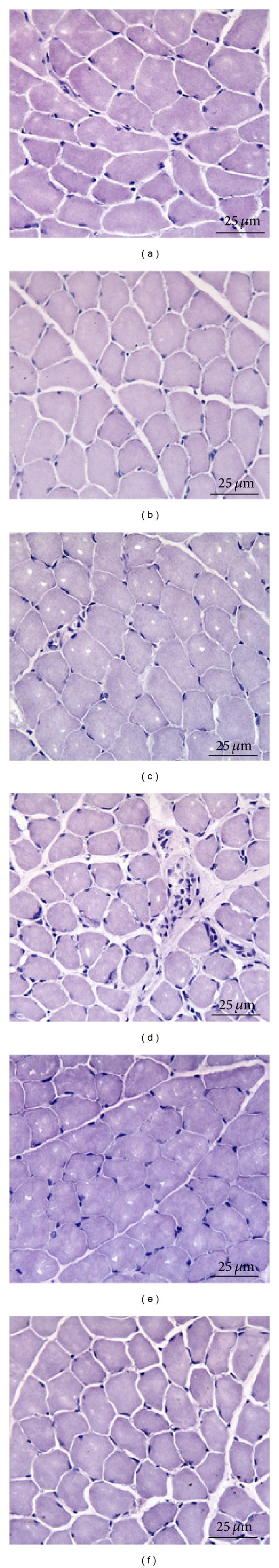
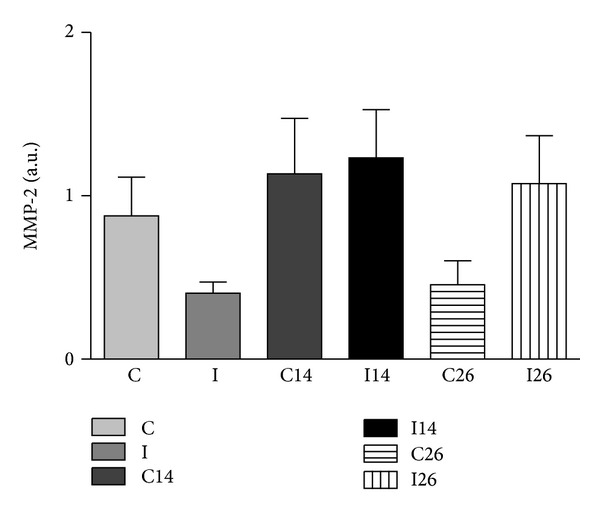
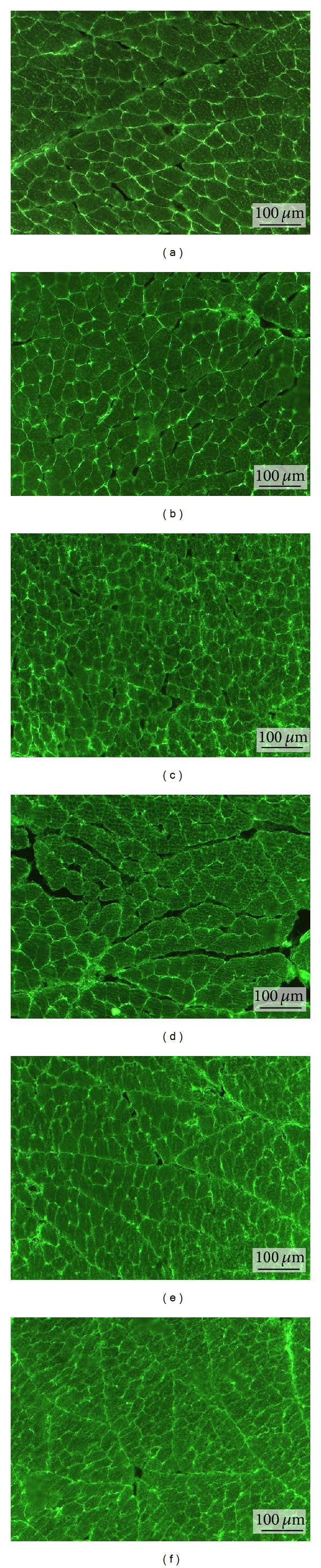
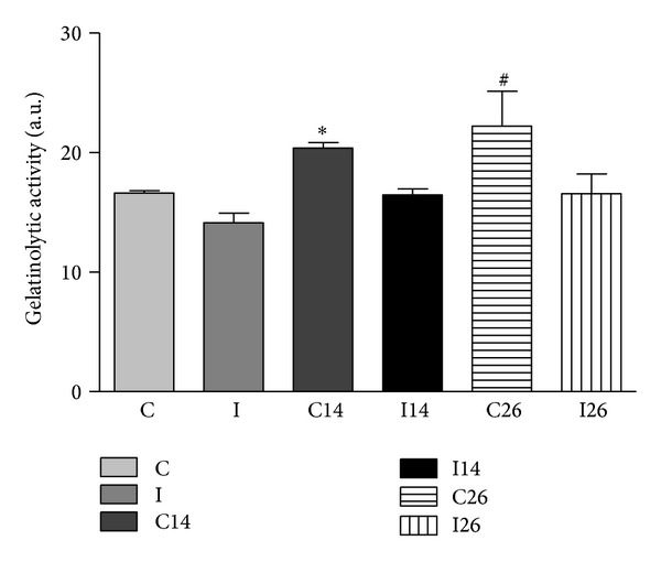
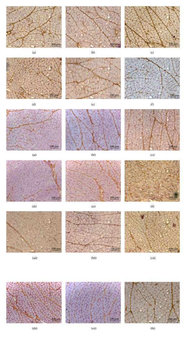
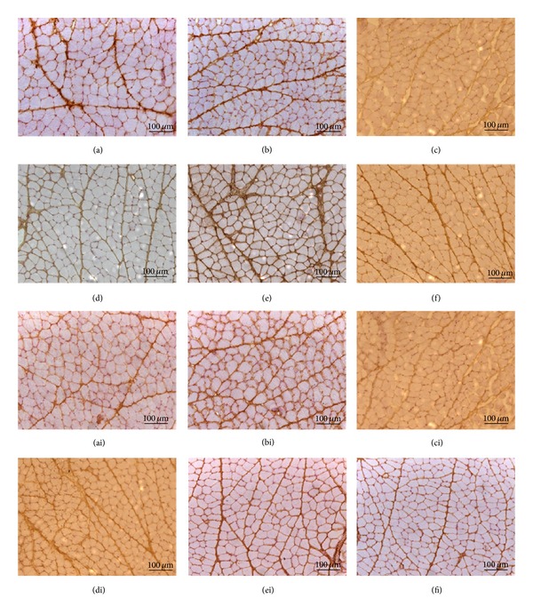
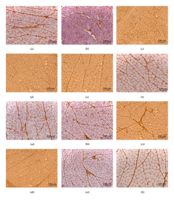
Although disorders of the stomatognathic system are common, the mechanisms involved are unknown. Our objective was to study the changes in the masseter muscles after unilateral exodontia. Molar extraction was performed on Wistar rats (left side), and the animals were sacrificed after either 14 or 26 days. The masseter muscle was processed for histological analysis, conventional and in situ zymography, and immunohistochemistry. The morphological analysis showed unique and specific characteristics for the experimental group. By conventional zymography no significant values of 72 kDa MMP-2 (P < 0.05) were found in both of the sides of masseter muscle after 14 and 26 days of unilateral extraction. The in situ zymography showed gelatinolytic activity on all deep masseter muscles, with significant increase on the contralateral side after 14 and 26 days (P < 0.05). The immunohistochemistry demonstrated greater expression of MMP-2 than MMP-9 and MMP-14 in all masseter muscles and there were few differences in the staining of 4 TIMPs. This knowledge about morphology and molecular masticatory muscle remodeling following environmental interventions can be used to develop clinically successful treatments.
Figures
Similar articles
-
Morphofunctional compensation of masseter muscles in unilateral posterior crossbite patients.Eur J Histochem. 2016 Jun 13;60(2):2605. doi: 10.4081/ejh.2016.2605. Eur J Histochem. 2016. PMID: 27349311 Free PMC article.
-
Identification of matrix metalloproteinases and their tissue inhibitors type 1 and 2 in human masseter muscle.Arch Oral Biol. 2000 Jun;45(6):431-40. doi: 10.1016/s0003-9969(00)00020-0. Arch Oral Biol. 2000. PMID: 10775672
-
Exodontia-induced muscular hypofunction by itself or associated to chronic stress impairs masseter muscle morphology and its mitochondrial function.Microsc Res Tech. 2019 May;82(5):530-537. doi: 10.1002/jemt.23196. Epub 2019 Feb 11. Microsc Res Tech. 2019. PMID: 30741445
-
Ultrastructural abnormalities of muscle spindles in the rat masseter muscle with malocclusion-induced damage.Histol Histopathol. 2002 Jan;17(1):45-54. doi: 10.14670/HH-17.45. Histol Histopathol. 2002. PMID: 11813885
-
Zymographic techniques for the analysis of matrix metalloproteinases and their inhibitors.Biotechniques. 2005 Jan;38(1):73-83. doi: 10.2144/05381RV01. Biotechniques. 2005. PMID: 15679089 Review.
Cited by
-
Intracerebroventricular Injection of Alarin Increased Glucose Uptake in Skeletal Muscle of Diabetic Rats.PLoS One. 2015 Oct 6;10(10):e0139327. doi: 10.1371/journal.pone.0139327. eCollection 2015. PLoS One. 2015. PMID: 26439383 Free PMC article.
-
[Alteration of metabolic characteristics on the masseter muscle fiber of unilateral chewing rats and its adenosine monophosphate activated protein kinase regulatory mechanism].Hua Xi Kou Qiang Yi Xue Za Zhi. 2017 Jun 1;35(3):258-263. doi: 10.7518/hxkq.2017.03.006. Hua Xi Kou Qiang Yi Xue Za Zhi. 2017. PMID: 28675009 Free PMC article. Chinese.
-
Functional magnetic resonance imaging evaluation of masticatory muscle dysfunction in unilateral exodontia rabbits.Dentomaxillofac Radiol. 2022 Jul 1;51(5):20220022. doi: 10.1259/dmfr.20220022. Epub 2022 May 4. Dentomaxillofac Radiol. 2022. PMID: 35466684 Free PMC article.
-
Morphofunctional compensation of masseter muscles in unilateral posterior crossbite patients.Eur J Histochem. 2016 Jun 13;60(2):2605. doi: 10.4081/ejh.2016.2605. Eur J Histochem. 2016. PMID: 27349311 Free PMC article.
-
Immunofluorescence Evaluation of Myf5 and MyoD in Masseter Muscle of Unilateral Posterior Crossbite Patients.J Funct Morphol Kinesiol. 2020 Nov 7;5(4):80. doi: 10.3390/jfmk5040080. J Funct Morphol Kinesiol. 2020. PMID: 33467295 Free PMC article.
References
-
- Imauchi Y, Sakamoto T, Abe K. A case of atrophy of the masticatory muscles due to a masticatory habit. European Archives of Oto-Rhino-Laryngology. 2002;259(10):551–553. - PubMed
-
- Schellhas KP. MR imaging of muscles of mastication. The American Journal of Roentgenology. 1989;153(4):847–855. - PubMed
-
- Hunt N, Shah R, Sinanan A, Lewis M. Northcroft Memorial Lecture 2005: muscling in on malocclusions: current concepts on the role of muscles in the aetiology and treatment of malocclusion. Journal of Orthodontics. 2006;33(3):187–197. - PubMed
-
- Cao Y, Xie QF, Yang ZH. Electrical pain thresholds of face and finger in patients with chronic masticatory muscle pain. Zhonghua Kou Qiang Yi Xue Za Zhi. 2009;44:373–376. - PubMed
-
- Kjaer DK, Sørensen TH, Knudsen HJ. Decreased fetal movements and massive fetomaternal transfusion. Ugeskr Laeger. 2004;166:3826–3827. - PubMed
Publication types
MeSH terms
Substances
LinkOut - more resources
Full Text Sources
Other Literature Sources
Molecular Biology Databases
Miscellaneous
